
Abstract
Breast cancer is one of the most common types of cancer worldwide. However, early diagnosis and detection by mammography contribute significantly to reducing its mortality. In this context, a key aspect within quality assurance programs is the measurement of radiation dose, which is essential to optimize the technique. The dosimetric magnitude that best characterizes the carcinogenic risk induced by ionizing radiation in mammography is the mean glandular dose (MGD). To establish the Diagnostic Reference Levels (DRLs) in mammography, adequate management of DGM is crucial. This process should begin with the promotion of a safety culture, the training of personnel, the assignment of responsibilities and the implementation of tools and methods for their evaluation. In addition, the success of this strategy requires the support of adequate regulation and sustainable financing by the competent authorities. Therefore, mammography services must adopt radiation protection measures through the implementation of DRLs. A methodology for establishing DRLs in mammography is presented, addressing their importance, the dosimetric magnitudes to be considered, and the data collection process, with the aim of defining and applying these reference levels effectively.
Keywords
breast cancer, mammography, mean glandular dose, reference levels for diagnosis
INTRODUCTION
Breast cancer is one of the most common types of cancer in women. In 2020, it was the most common cancer globally, although it ranked fifth among the leading causes of cancer death. However, early diagnosis and detection through mammography exams contribute significantly to reducing breast mortality (WHO, 2022). A key factor in the fight against breast cancer is the economic aspect. In high-income countries, the prognosis for patients is usually favorable, while in low- or middle-income countries, the reality is very different. Lack of access to early diagnosis and timely treatment is the main cause of this inequality. In 2020, almost three-quarters of breast cancer deaths worldwide occurred in low- and middle-income countries (IARC, 2021). The incidence and mortality of breast cancer in Latin American women shows the same behavior as globally. The incidence has been increasing in the last two decades, ranking among the first or second type of cancer in women depending on the country (Mora et al., 2014).
Currently, mammography is considered the best tool for the early detection of breast cancer, playing a fundamental role in both the diagnosis and localization of lesions in biopsy and therapy procedures. Among the pathological signs associated with breast cancer are calcifications, nodules, distortions, and asymmetries. The detection of these lesions in a mammogram can be difficult, since their attenuation or absorption of X-rays is very similar to that of healthy tissues. This is because mammography projects the volume of the breast in a two-dimensional (2D) image, which can make it difficult to differentiate between normal and pathological structures (Castillo et al., 2015). Despite its effectiveness in breast cancer screening, mammography has been the subject of debate due to the possible induction of breast cancer or alterations in nearby organs, such as the thyroid and lens (Sechopoulos et al., 2008; Pérez et al., 2022).
The effectiveness of a mammographic procedure depends primarily on its diagnostic quality, which necessitates the implementation of a quality assurance program to optimize its efficiency and safety (Milano et al., 2000). These protocols enable the acquisition of high-quality diagnostic images with a reasonable radiation dose, in accordance with the ALARA principle. Within a quality assurance program, one fundamental test is the measurement of radiation dose, a key aspect for the optimization of the technique. The dosimetric quantity that best characterizes the carcinogenic risk induced by ionizing radiation in mammography is the mean glandular dose (DGM) (European Commission, 2006).
The European Commission (2013) has established acceptable and achievable DGM values based on compressed breast thickness, which ranges from 21 to 90 mm for screening examinations, see Table 1. However, it is essential to consider individual differences among patients, such as breast thickness and glandular density, which can influence the required radiation dose and exposure time. Likewise, the presence of breast implants increases the number of required projections and the thickness of breast tissue, making it necessary to adjust the technique to ensure 1 complete and interpretable study (Chetlen et al., 2016).
PMMA thickness (mm) | Equivalent breast thickness (mm) | Maximum DGM (mGy) | Maximum DGM (mGy) |
|---|---|---|---|
[empty cell] | [empty cell] | Acceptable value | Achievable value |
20 | 21 | 1,0 | 0,6 |
30 | 32 | 1,5 | 1,0 |
40 | 45 | 2,0 | 1,6 |
45 | 53 | 2,5 | 2,0 |
50 | 60 | 3,0 | 2,4 |
60 | 75 | 4,5 | 3,6 |
70 | 90 | 6,5 | 5,1 |
PPMA: polymethyl methacrylate, an acrylic material with a density similar to that of the breast.
The concept of the diagnostic reference level (DRL) is internationally recognized as a key tool for optimizing patient exposure to radiation (ICRP, 2017). DRLs serve as guidance values for evaluating and adjusting the doses administered during diagnostic imaging procedures. Their implementation enables institutions and radiology services to systematically monitor, compare, and manage radiation exposure in radiological studies, thereby contributing to a safer and more efficient clinical practice.
Comparison of the DGM with national or international DRLs provides an overview of practice and facilitates its optimization. It is important to note that DRLs should not be interpreted as strict limits, but rather as dose indicators that allow identification of opportunities for improvement. A well-established DRL contributes to adjusting exposure when necessary, ensuring a balance between image quality and patient safety. According to the International Commission on Radiological Protection in its publication 135 (ICRP, 2017), a DRL is defined as the 75th percentile of the distribution of median values obtained in participating institutions through a survey.
In this context, a methodology is presented for establishing DRLs in mammography, addressing their importance, the dosimetric quantities to consider, and the data collection process, with the aim of effectively defining and applying these reference levels.
Justification for radiological examination in mammography.
The first step in effective management of radiation dose in mammography is to ensure that each examination is performed only for duly justified clinical indications. Any request in which mammography has a high probability of providing relevant diagnostic information for early detection or evaluation of breast disease is considered appropriate. In cases where such information can be obtained accurately and in a timely manner using techniques that involve a lower radiation dose or that do not use ionizing radiation, such as magnetic resonance imaging or breast ultrasound, mammography should not be the method of choice.
One appropriate justification, accompanied by optimized practice, makes it possible to avoid unnecessary repetition of studies and, consequently, unjustified exposure to radiation. It also contributes to maintaining a systematic record of the examinations performed and to preserving monthly data, fundamental for the estimation and control of the DGM, thus promoting continuous improvement in radiological practice.
Optimization of radiological practice.
Since its inception, mammography has been the reference technique for the early detection of breast cancer. With the development of digital mammography and the incorporation of tomosynthesis, the early detection rate has significantly improved. However, the risk associated with radiation exposure, especially the potential for carcinogenesis, remains a major concern, particularly in population-based screening programs aimed at women over 40 years of age. In this context, the optimization and continuous monitoring of the administered dose are essential to ensure safe and effective practice.
A key measure in this process is the periodic performance of quality controls of the mammography equipment, with the aim of ensuring that the administered dose is the minimum necessary to obtain images with adequate diagnostic quality. The mammary gland is composed of adipose tissue and glandular tissue, the latter being the most radiosensitive and where cancer can originate. For this reason, the parameter used to evaluate the absorbed dose in mammography is the DGM. The precise estimation of the DGM presents challenges, since the radiation distribution is not uniform: glandular tissues receive different amounts of radiation depending on the angle of incidence of the beam and the depth it reaches. Furthermore, factors such as equipment configuration, the acquisition techniques used, and patient characteristics significantly influence the administered dose.
Optimization of mammographic practice is supported by various reference sources, including the American College of Radiology (ACR, 1999), the European Community Guidelines (European Commission, 2013), Report 149 of the National Council on Radiation Protection (NCRP, 2004), and the technical document of the International Atomic Energy Agency (IAEA-TECDOC-1517, 2006). Avoiding unnecessary studies not only reduces radiation exposure, but also minimizes costs, avoids incidental findings without clinical relevance, and reduces anxiety in patients.
Several studies have demonstrated the usefulness of DRLs as a key tool in radiation protection, allowing the establishment of indicative thresholds that help optimize patient exposure without compromising the diagnostic value of the study (Hart et al., 2012). However, in some Latin American countries, the implementation of DRLs has been limited due to outdated regulations governing the use of ionizing radiation in medicine. In this regard, as part of the OIEA program on Radiation Protection of Patients in Medical Exposures (TSA3), Mora and colleagues (2014) conducted a survey in 13 Latin American countries, establishing DRL values in mammography. However, institutional participation was limited in most countries.
In Chile, Leyton et al. (2015) estimated the DGM in 6 digital mammography systems and proposed preliminary DRL values using PMMA phantoms. In Colombia, Amaya y Muñoz (2021) determined DRLs in conventional radiology, mammography, tomography, and fluoroscopy practices. In Brazil, starting in 2022, the project “Niveles de Referencia Diagnósticos en Latinoamérica” (NRDLatin, 2022) was implemented, which enabled a platform for the systematic collection of technical data from radiological examinations. Its objective is to establish strategies to define DRLs by anatomical region and clinical indication, in both tomography and radiography and mammography. This project includes the preparation of a report with recommendations based on the guidelines of the OIEA and the OMS, as well as a national and regional document, under the supervision of the Colegio Brasileño de Radiología. The responsibility for complying with the study methodology lies with each participating institution. As a result, each center will be confidentially provided with the local typical value, the national DRL value (stratified by clinical indication, body mass index, and equipment technology), along with suggestions to improve protocols, training through workshops, audiovisual materials, and the official publication of the DRLs at the national and Latin American level.
Estimation of the mean glandular dose in mammography.
There are two formalisms for the calculation of DGM in mammography: the Dance model (European model) and the Wu and Boone models (North American models).
The Dance model (Dance et al., 2000; Dance, Young, van Engen, 2009) is a simple model based on a fixed semicircular cross-section cylinder, with a diameter of 160 mm. Typical glandularity values have been provided for women in two age groups: 40-49 years and 50-64 years. DGM is determined by the following equation:
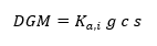
(1)
where: , represents the incident air kerma at the upper surface of the breast; , is a factor that depends on the value of the half-value layer (HVL) and the breast thickness; , a factor that accounts for the percentage of glandularity of the breast; and , a factor that depends on the anode/filter combination.
This model was introduced in the United Kingdom in 1989 through a standard protocol for dosimetry in conventional projection mammography (IPSM 1989). Subsequently, it was extended (IPEM 2005) to include the use of X-ray spectra with different anode/filter combinations (through the introduction of factor s) and to provide dosimetry in a variety of breast glandularities (with the incorporation of factor c). A similar methodology has been adopted in European protocols (CE 1996, 2006, and 2013) and by the OIEA (2007). These protocols use conversion factors based on Monte Carlo calculations (Dance et al., 2000; Dance, Young, van Engen, 2009) to relate the measurements to the DGM.
The Wu model (Sobol and Wu, 1997) is a simple model based on a cylinder with a semi-elliptical cross-section. To estimate the DGM, this model uses the following equation:
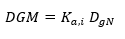
(2)
where: the first term has already been defined, and the second term is the glandular dose normalized per unit.
The DGM depends on the anode/filter combination and the glandularity of the breast. For this calculation, it is assumed that the breast is composed of 50% glandular tissue and 50% adipose tissue. The Wu model provides the parameterization equations to calculate the DGM for different anode/filter combinations and different glandularities (Sobol and Wu, 1997). However, this method is limited to three specific spectra: Mo/Mo, Mo/Rh, and Rh/Rh, so its application is restricted to mammography systems manufactured by the company General Electric (Samara, Tsapakib, Srameck, 2019).
The Boone model (Boone, 1999; Boone, 2002) is an extension of the Wu model, based on a circular cross-section. This model uses monoenergetic X-rays up to 120 keV and supports a greater number of anode/filter combinations, including W/Rh and W/Ag. Unlike its predecessors, the Boone model provides a better description of energy absorption in the central homogeneous region of the breast, considering both adipose and glandular tissue. To estimate the DGM, this model uses equation (2).
DRL values in mammography.
DRLs in mammography are a key tool for monitoring and optimizing radiation doses, allowing comparison between countries, institutions, and mammography units.
Although various guidelines and recommendations have provided clarity on the methodologies used in establishing DRLs in mammography, challenges still exist in their standardization. Among the most relevant considerations are:
Data source: it is recommended that information derived from patients, rather than data obtained from phantoms, be used to establish DRLs.
The protocol: it is suggested that DRLs be stratified according to breast thickness and detector technology, and that the median, rather than the mean, be used for their calculation.
The percentiles: DRLs should be established at the 75th percentile, with a minimum sample size of 50 patients.
Despite these recommendations, the lack of a standardized method for estimating DGM continues to hinder comparison between studies conducted in different mammography systems.
Establishment of DRLs in Mammography.
(a) Regulation.
To ensure adequate practice in mammography, it is essential to have a regulatory framework that establishes the DRL values, as well as their correct application and the optimization of radiation protection in medical exposures. Given that the management of the dose delivered to patients varies significantly between countries (Martín et al., 2013), it will be necessary to apply a creative and flexible approach both for the determination of DRLs and for the implementation of optimization programs. It is essential that these strategies are adapted to the particularities of each national context, in order to guarantee safe and effective radiological practice aligned with the principles of radiation protection.
(b) Selection of the dosimetric quantity.
The selected dosimetric quantity must be directly related to the imaging modality studied. Table 2 presents the recommended dosimetric quantities for establishing DRLs in mammography, as indicated by the ICRP in its Publication 135 (ICRP, 2017).
Table 2. Appropriate dosimetric quantities for establishing DRLs in mammography.
Modality | Recommended magnitude | Recommended unit |
|---|---|---|
Mammography | Ka,i Ka,e DGM | mGy mGy mGy |
Adapted from Reference (ICRP, 2017).
Although air kerma is relatively easy to measure and useful for making quick comparisons, it is neither additive nor representative of the actual risk to the patient. In contrast, the DGM, although it cannot be measured directly (since it must be estimated from simple measurements and the use of conversion factor tables), is the most representative quantity of the risk associated with mammographic exposure.
(c) Values of DRLs.
DRLs can be established from the distribution of the medians of the dosimetric quantities measured in one sample of individual patients or equipment (mammography, tomosynthesis) in different geographic regions. The median is considered a more robust statistical estimator than the mean, since it is less sensitive to outliers and, with a larger volume of dosimetric data, provides a more reliable representation of the patient population.
For areas with between 10 and 20 mammography rooms, the local DRL should be established as the third quartile (75th percentile) of the distribution of the medians of the measured doses. In cases where a small number of rooms is available or one single institution is involved, a “typical value” may be defined as the median of the recorded doses, which can be used for purposes similar to those of a local DRL. Establishing DRLs at the national level requires a representative sample, approximately 30% of institutions (hospitals, clinics) distributed throughout the country. Regional DRLs, for their part, apply to groups of countries that share similar radiological practices (ICRP, 2017).
(d) Facilities.
To establish DRLs, it is crucial to delimit the geographical area in which they will be evaluated and subsequently applied. In a local setting, a DRL can be derived from a group of 10 to 20 mammography rooms. However, in large countries with hundreds of health centers, conducting an exhaustive sampling would be a complicated task. Results from 20 to 30 rooms may be adequate initially, provided that a sufficient number of patients (≥ 50) is included. In smaller countries with fewer than 50 rooms, one initial sampling covering 30 to 50% of these might be sufficient. As data collection infrastructure improves, consideration can be given to expanding the number of rooms or institutions included in subsequent studies to achieve a more representative coverage.
Once the DRLs have been established, optimization studies with an interval of three (3) years can be considered. If continuous data collection is possible, the dose management process can take the form of a periodic review.
(e) Patients or phantoms.
Most procedures for establishing DRLs are based on measurements performed on individual patients, classified by variables such as age, weight, and height. It is recommended to include at least 50 patients per mammography room, preferably restricting the range of compressed breast thicknesses to ensure sample homogeneity. However, in certain circumstances, phantoms can be useful for evaluating the technical performance of equipment. Nevertheless, phantoms should not be considered a substitute for surveys based on real patient examinations, as they do not accurately reflect real clinical conditions.
(f) Data collection methods.
Manual data collection remains an option, particularly in prospective studies. When the number of mammography rooms is small, printed forms specifically adapted to the type of examination may be used. However, the implementation of automatic exposure control systems in the context of DRLs offers important advantages, including the possibility of conducting retrospective reviews of the information recorded during patient examinations.
Data collection through the Radiology Information System (RIS) allows the inclusion of a significantly larger number of patients, thereby improving the representativeness of the data. In this context, the DICOM standard (Digital Imaging and Communication in Medicine) has developed a specific format for recording dosimetric information, the RDSR (Radiation Dose Structured Report), designed to store detailed information about radiation dose across various imaging modalities (DICOM, 2007). DICOM images are automatically transmitted from the PACS system to software that interprets DICOM headers, recording quantities such as Ka,i and DGM.
Automatic dose management also enables quick access to key data, such as patient age and weight, doses received, and technical parameters of the equipment. It also facilitates the export of filtered data sets for subsequent analysis.
Application of DRLs in mammography.
To ensure the correct application of DRLs in mammography, the following steps are recommended:
Collection of patient data, either manually or through electronic systems.
Estimation or calculation of the DGM per patient in each ward or institution.
Calculation of the median DGM, which is considered the typical dose value.
Comparison of the median with the DRLs (if established values are available): if the median is less than the DRL, return to step (i) and verify the validity of the data; if the median is greater than the DRL, continue with the next step.
Review of the technique used, exposure parameters, and equipment performance.
Verification that image quality is not compromised during the optimization process.
Recommendation and implementation of a practice optimization strategy based on the findings obtained.
Bibliografía
Amaya, E. & Muñoz, E. (2021). Determinación de los niveles de referencia de dosis (DRL) para diagnóstico de baja y media complejidad en Servicios Especiales de Salud Hospital Universitario de Caldas de Colombia (SES-HUC). Revista Investigaciones y Aplicaciones Nucleares, 5, 84-98. doi:10.32685/2590-7468/invapnuclear.5.2021.604
American College Radiology. Committee on Quality Assurance in Mammography. Medical Physicist’s Section. In Mammography Quality Control Manual 1999. (ACR, Washington, D.C), 1999.
Boone, J. M. (1999). Glandular breast dose for monoenergetic and high-energy X-ray beams: Monte Carlo assessment. Radiology 213(1), 23-37.
Boone, J. M. (2002). Normalized glandular dose (DgN) coefficients for arbitrary X-ray spectra in mammography: computer-fit values of Monte Carlo derived data. Med. Phys. 29(5), 869-875.
Breast Cancer Awareness Month 2021 - IARC Cancer. Recuperado de http://www.iarc.who.int/feature-news/breast-cancer-awareness-month-2021/
Castillo, M., Chevalier, M., et al (2015). Estudio de los valores de dosis a pacientes en exámenes de tomosíntesis de mama estimados con dos métodos distintos. Rev. Fis. Med. 16(3), 35-46.
Chetlen, A. L., Brown, K. L., et al (2016). Scatter radiation dose from digital screening mammography measured in a representative patient population. Am. J. Roentgenol. 206, 359-65.
Commission of the European Communities. European guidelines for quality assurance in breast cancer screening and diagnosis, 4th edition. European communities (2006). ISBN 92-79-01258-4.
Dance, D. R., Skinner, C. L., et al (2000). Additional factors for the estimation of mean glandular breast dose using the UK mammography dosimetry protocol. Phys. Med. Biol. 45(11), 3225-3240.
Dance, D. R., Young, K. C. & van Engen, R. E. (2009). Further factors for the estimation of mean glandular dose using the United Kingdom, European and IAEA breast dosimetry protocols. Phys. Med. Biol. 54(14), 4361-4372.
Digital Imaging and Communications in Medicine (DICOM). Supplement 127: CT Radiation Dose Reporting (Dose SR). DICOM Standards Committee, 2007.
European Commission, Directorate-General for Health and Consumers, 2013. Karsa, L., Holland, R., Broeders, M., Wolf, C., Perry, N. & Törnberg S. (eds). European guidelines for quality assurance in breast cancer screening and diagnosis 4th edition, supplements.
Hart, D., Hillier, M. C. & Shrimpton, P. C. (2012). Doses to Patients from Radiographic and Fluoroscopic X-Ray Imaging Procedures in the UK? 2010 review. Chilton, Health Protection Agency, Centre for Radiation, Chemical and Environmental Hazards, Radiation Protection Division.
IAEA-TECDOC-1517, Control de Calidad en Mamografía. Editado por OIEA. Viena 2006.
ICRP 2017. Diagnostic reference levels in medical imaging. ICRP publication 135. Ann. ICRP 46.
Leyton, F., Nogueira, M., Dantas, M., Duran, M. P. & Ubeda, C. (2015). Mean glandular dose in six digital mammography services in Santiago, Chile: Preliminary Reference Levels. Radiat. Prot. Dosim., 165 (1-4), 115-120. doi.10.1093/rpd/ncv092
Martin, C. J., Le Heron, J., Borrás, C., Sookpeng, S. & Ramírez, G. (2013). Approaches to aspects of optimization of protection in diagnostic radiology in six continents. J. Radiol. Prot., 33(4), 711-34.
Milano, F., Maggi, E. & Roselli Del Turco, M. (2000). Evaluation of the effect of a quality control programme in mammography on technical and exposure parameters. Radiat. Prot. Dosim. 90, 263-266.
Mora, P., Blanco, S., et al (2014). Latin American dose survey results in mammography studies under IAEA programme: radiological protection of patients in medical exposures (TSA3). Radiat. Prot. Dosim., 163(4), 473-479. doi:10.1093/rpd/ncu205
NCRP Report No. 149. A guide to mammography and other breast imaging procedures (2004).
NRD Brasil e América Latina, NRDLatin 2022. Niveles de Referencia de Diagnóstico en Latinoamérica. Recuperado de http://www.nrdamericalatina.com.br
Pérez, J. A., Roldán, V. S., Gordillo, A. K., Mena, A. F., Brito, S. & Soteldo, C. (2022). Dosis glandular promedio en glándula mamaria y dosis de radiación en glándula tiroides y cristalino en mujeres con y sin implantes mamarios durante diferentes modalidades mamográficas. Radiología 64, 11-19. doi.org/10.1016/j.rx.2020.10.013
Sechopoulos, I., Suryanarayanan, S., et al (2008). Radiation Dose to Organs and Tissues from Mammography: Monte Carlo and Phantom Study. Radiology 246, 434-443.
Sobol, W. T. & Wu, X. (1997). Parametrization of mammography normalized average glandular dose tables. Med. Phys. 24(4), 547-554.
Samara, E. T., Tsapakib, V. & Sramekc, D. (2019). Dose management software implementation in mammography. Physica Medica, 68, 88-95.
World Health Organization 2022 Cancer. Recuperado de http://ww.who.int/mediacentre/factsheets/fs297/en/
Declarations
Funding
No financing.
Conflict of interest
None.
Authorship contributions
Drafting – original draft: Manuel A.Villarreal U., Resumen, Abstract.
Writing–review and editing: Manuel A.Villarreal U., Resumen, Abstract.