

Abstract
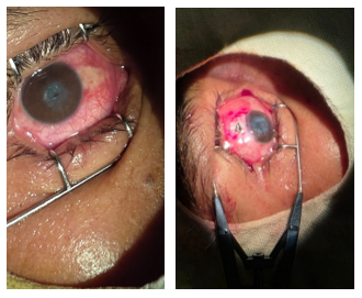
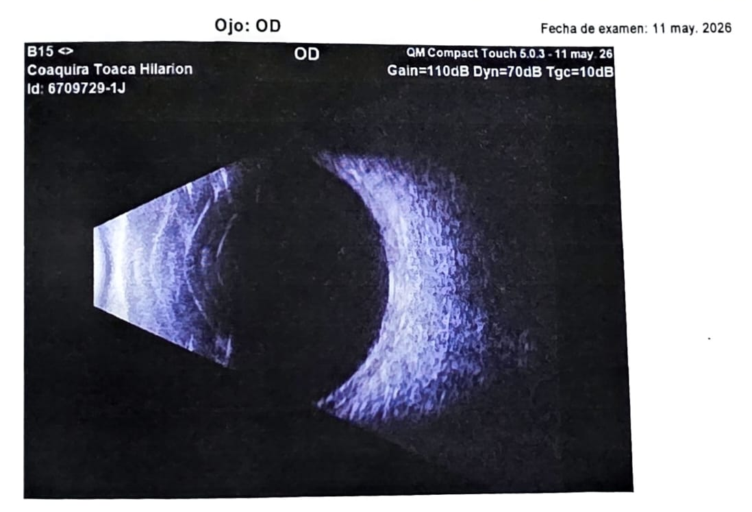
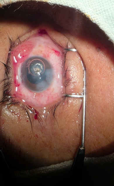
Introduction: Open globe trauma with an intraocular foreign body constitutes an ophthalmological emergency that irreversibly threatens vision. Its prognosis depends on the location of the injury, the structures involved, and the speed of treatment. Case report: A 34-year-old male patient, with no relevant medical history, presented with right ocular trauma from a metallic foreign body, eight hours prior, with intense pain, tearing, and sudden vision loss. Biomicroscopy revealed a 4 mm penetrating corneal wound, a positive Seidel sign, a narrowed anterior chamber, an intralenticular foreign body, and a traumatic cataract. Ultrasound ruled out retinal and vitreous detachment. Corneal repair, extraction via a scleral tunnel, and aspiration of the lens nucleus were performed; intraocular lens implantation was deferred. Discussion. The corneal location of the wound and the anterior position of the foreign body define a favorable prognostic stratum. Its proximity to the endothelium determined the extraction route and the postponement of aphakic correction. Conclusions: In cases of penetrating ocular trauma, preserving vision depends not only on acting quickly but also on correctly deciding what needs to be addressed first. The presence of a metallic foreign body, a corneal wound, and a traumatic cataract necessitates simultaneous consideration of the integrity of the eyeball. The decision not to immediately implant an intraocular lens in a recently injured eye may represent a prudent reconstructive strategy. In occupational cases, workplace eye protection is identified as the most effective intervention.
Keywords
Ocular trauma, open globe, intraocular foreign body, traumatic cataract, lensectomy
CONCLUSIONES
El caso pone de manifiesto que, frente a un trauma ocular penetrante, preservar la visión no depende únicamente de actuar con rapidez, sino de decidir correctamente qué debe resolverse primero. La presencia de un cuerpo extraño metálico, una herida corneal y una catarata traumática obliga a considerar simultáneamente la integridad del globo, el riesgo infeccioso, la viabilidad corneal y las posibilidades futuras de rehabilitación visual. La decisión de no implantar inmediatamente una lente intraocular puede parecer, desde una perspectiva exclusivamente refractiva, una postergación del tratamiento; sin embargo, en un ojo recientemente lesionado puede representar una estrategia reconstructiva prudente. El objetivo de la primera intervención no siempre es devolver inmediatamente la agudeza visual, sino conservar las condiciones anatómicas que permitan recuperarla posteriormente. El origen laboral del accidente señala, el punto donde la intervención resulta más eficiente. Ninguna técnica quirúrgica compensa la ausencia de protección ocular en el puesto de trabajo y la notificación sistemática de estos casos constituye el insumo mínimo para sostener políticas de prevención sobre todo en países sin registros consolidados de trauma ocular ocupacional.
REFERENCIAS BIBLIOGRÁFICA
Kuhn F, Morris R, Witherspoon CD. Birmingham Eye Trauma Terminology (BETT): terminology and classification of mechanical eye injuries. Ophthalmol Clin North Am. 2002;15(2):139-43. https://doi.org/10.1016/S0896-1549(02)00004-4
Blanch RJ, McMaster D, Patterson TJ. Management of open globe injury: a narrative review. Eye (Lond). 2024;38(16):3047-51. https://doi.org/10.1038/s41433-024-03246-3
Loporchio D, Mukkamala L, Gorukanti K, Zarbin M, Langer P, Bhagat N. Intraocular foreign bodies: a review. Surv Ophthalmol. 2016;61(5):582-96. https://doi.org/10.1016/j.survophthal.2016.03.005
Cai M, Zhang J. Epidemiological characteristics of work-related ocular trauma in southwest region of China. Int J Environ Res Public Health. 2015;12(8):9864-75. https://doi.org/10.3390/ijerph120809864
Jabłoński M, Winiarczyk M, Biela K, Bieliński P, Jasielska M, Batalia J, et al. Open globe injury (OGI) with a presence of an intraocular foreign body (IOFB): epidemiology, management, and risk factors in long term follow-up. J Clin Med. 2023;12(1):190. https://doi.org/10.3390/jcm12010190
Zhang Y, Zhang M, Jiang C, Qiu HY. Intraocular foreign bodies in China: clinical characteristics, prognostic factors, and visual outcomes in 1,421 eyes. Am J Ophthalmol. 2011;152(1):66-73.e1. https://doi.org/10.1016/j.ajo.2011.01.014
Greven CM, Engelbrecht NE, Slusher MM, Nagy SS. Intraocular foreign bodies: management, prognostic factors, and visual outcomes. Ophthalmology. 2000;107(3):608-12. https://doi.org/10.1016/S0161-6420(99)00134-7
Vingopoulos F, Wang Y, Grob S, Li CYL, Eliott D, Kim LA, et al. Open globe injury with intraocular foreign body. J Vitreoretin Dis. 2021;5(4):288-94. https://doi.org/10.1177/2474126420965033
Patel SN, Langer PD, Zarbin MA, Bhagat N. Diagnostic value of clinical examination and radiographic imaging in identification of intraocular foreign bodies in open globe injury. Eur J Ophthalmol. 2012;22(2):259-68. https://doi.org/10.5301/EJO.2011.8347
Bhagat N, Nagori S, Zarbin M. Post-traumatic infectious endophthalmitis. Surv Ophthalmol. 2011;56(3):214-51. https://doi.org/10.1016/j.survophthal.2010.09.002
Ahmed Y, Schimel AM, Pathengay A, Colyer MH, Flynn HW Jr. Endophthalmitis following open-globe injuries. Eye (Lond). 2012;26(2):212-7. https://doi.org/10.1038/eye.2011.313
Tabatabaei SA, Rajabi MB, Tabatabaei SM, Soleimani M, Rahimi F, Yaseri M. Early versus late traumatic cataract surgery and intraocular lens implantation. Eye (Lond). 2017;31(8):1199-204. https://doi.org/10.1038/eye.2017.57
Sen P, Shah C, Sen A, Jain E, Mohan A. Primary versus secondary intraocular lens implantation in traumatic cataract after open-globe injury in pediatric patients. J Cataract Refract Surg. 2018;44(12):1446-53. https://doi.org/10.1016/j.jcrs.2018.07.061
Loporchio D, Mukkamala L, Gorukanti K, Zarbin M, Langer P, Bhagat N. Intraocular foreign bodies: a review. Surv Ophthalmol. 2016;61(5):582-596. https://doi.org/10.1016/j.survophthal.2016.03.005
Blanch RJ, McMaster D, Patterson TJ. Management of open globe injury: a narrative review. Eye (Lond). 2024;38(16):3047-3051. https://doi.org/10.1038/s41433-024-03246-3
Shah M, Shah S, Shashank S, Prasad V, Parikh A. Visual recovery and predictors of visual prognosis after managing traumatic cataracts in 555 patients. Indian J Ophthalmol. 2011;59(3):217-222. https://doi.org/10.4103/0301-4738.81043
Declaraciones
Financiación
Los autores no recibieron financiación para el desarrollo de la presente investigación.
Conflictos de interés
Los autores declaran que no existe conflicto de intereses.
Contribución de autoría
Conceptualización: Dilma Villca Villca.
Curación de datos: Jaykel Evelio Gómez Triana
Análisis formal: Rolando Quispaya Quispe
Investigación: Dilma Villca Villca.
Metodología: Dilma Villca Villca.
Administración del proyecto: Dilma Villca Villca
Recursos: Freddy Ednildon Bautista-Vanegas.
Software: Jorge Marquez Molina.
Supervisión: Freddy Ednildon Bautista-Vanegas.
Validación: Jaykel Evelio Gómez Triana.
Visualización: Freddy Ednildon Bautista-Vanegas.
Redacción – borrador original: Dilma Villca Villca, Rolando Quispaya Quispe, Carlos Yohan Cruz Martínez, Freddy Ednildon Bautista-Vanegas, Jaykel Evelio Gómez Triana, Jorge Marquez Molina, Amanda Soto Martorell.
Redacción – revisión y edición: Dilma Villca Villca, Rolando Quispaya Quispe, Carlos Yohan Cruz Martínez, Freddy Ednildon Bautista-Vanegas, Jaykel Evelio Gómez Triana, Jorge Marquez Molina, Amanda Soto Martorell.