

Abstract
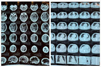
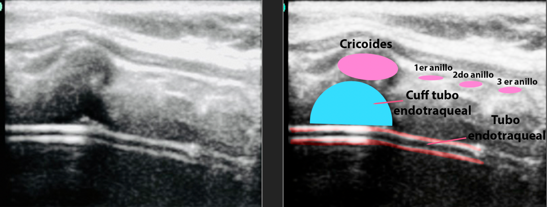
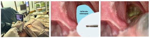
Introduction: Percutaneous dilation tracheostomy has largely replaced surgical tracheostomy in most intensive care units. Bronchoscopy remains the standard guidance method, although it partially obstructs the tube lumen, increases carbon dioxide levels, and requires equipment that is not always available. At high altitudes, where arterial oxygen levels are already reduced, these disadvantages are even more significant. Case report: An 82-year-old male patient, a permanent resident at 3650 meters above sea level, was admitted to the intensive care unit for a right hemisphere ischemic stroke, hemodynamic and ventilatory instability, atrial flutter, bilateral pleural effusion, and hospital-acquired pneumonia. After 16 days of invasive ventilation, a bedside tracheostomy was planned. A 5-10 MHz linear transducer identified the thyroid and cricoid cartilages, the tracheal rings, the pretracheal tissue, and the absence of vessels along the planned tracheostomy path. The videolaryngoscope allowed for the withdrawal of the endotracheal tube under direct vision until the cuff was positioned immediately below the vocal cords. The cannula was inserted using a modified Seldinger technique, with oxygenation goals appropriate for high altitude. Discussion: Ultrasound and videolaryngoscopy address different problems: the former defines the extraluminal anatomy of the anterior neck, while the latter monitors the endolaryngeal position of the tube and its cuff. Their combination reproduces much of the information provided by bronchoscopy without inserting an instrument into the tube lumen, a significant advantage in a patient whose baseline arterial oxygen pressure is approximately 56 mmHg. Conclusions: Ultrasound-guided, videolaryngoscopy-assisted percutaneous tracheostomy by dilation proved feasible at the bedside of a neurocritically ill patient residing at 3650 meters above sea level, without immediate complications and respecting the oxygenation goals specific to high altitude.
Keywords
Percutaneous tracheostomy, POCUS, videolaryngoscopy, high altitude, intensive care, bronchoscopy
CONCLUSIONES
La traqueostomía percutánea por dilatación guiada por ecografía y asistida por videolaringoscopia resultó factible a la cabecera de un paciente neurocrítico residente a 3650 msnm, sin complicaciones inmediatas y respetando las metas de oxigenación propias de la gran altitud. La utilidad de la combinación proviene de la complementariedad entre ambas herramientas, la ecografía define la anatomía que la palpación no discrimina y el videolaringoscopio vigila el momento en que el tubo endotraqueal deja de estar protegido. Frente a la broncoscopia, la diferencia práctica más relevante no es la calidad de la imagen sino la conservación de la luz del tubo, condición que adquiere otro significado cuando la presión arterial de oxígeno basal del paciente se sitúa cerca de 56 mmHg. La relevancia del caso clínico justifica que las UCI situadas en la altitud, con acceso irregular a broncoscopia flexible, consideren esta estrategia dentro de un protocolo con registro sistemático de variables gasométricas y de complicaciones, único camino para convertir una observación aislada en evidencia utilizable.
References
- 1 Shelden CH, Pudenz RH, Freshwater DB, Crue BL. A new method for tracheotomy. J Neurosurg. 1955;12(4):428-31. https://doi.org/10.3171/jns.1955.12.4.0428
- 2 Ciaglia P, Firsching R, Syniec C. Elective percutaneous dilatational tracheostomy. A new simple bedside procedure; preliminary report. Chest. 1985;87(6):715-9. https://doi.org/10.1378/chest.87.6.715
- 3 Delaney A, Bagshaw SM, Nalos M. Percutaneous dilatational tracheostomy versus surgical tracheostomy in critically ill patients: a systematic review and meta-analysis. Crit Care. 2006;10(2):R55. https://doi.org/10.1186/cc4887
- 4 Cabrini L, Monti G, Landoni G, Biondi-Zoccai G, Boroli F, Mamo D, et al. Percutaneous tracheostomy, a systematic review. Acta Anaesthesiol Scand. 2012;56(3):270-81. https://doi.org/10.1111/j.1399-6576.2011.02592.x
- 5 Dennis BM, Eckert MJ, Gunter OL, Morris JA Jr, May AK. Safety of bedside percutaneous tracheostomy in the critically ill: evaluation of more than 3,000 procedures. J Am Coll Surg. 2013;216(4):858-65. https://doi.org/10.1016/j.jamcollsurg.2012.12.017
- 6 Hosokawa K, Nishimura M, Egi M, Vincent JL. Timing of tracheotomy in ICU patients: a systematic review of randomized controlled trials. Crit Care. 2015;19(1):424. https://doi.org/10.1186/s13054-015-1138-8
- 7 Hashimoto DM, Axtell AL, Auchincloss HG. Percutaneous tracheostomy. N Engl J Med. 2020;383(20):e112. https://doi.org/10.1056/NEJMvcm2014884
- 8 Bösel J, Niesen WD, Salih F, Morris NA, Ragland JT, Gough B, et al. Effect of early vs standard approach to tracheostomy on functional outcome at 6 months among patients with severe stroke receiving mechanical ventilation: the SETPOINT2 randomized clinical trial. JAMA. 2022;327(19):1899-909. https://doi.org/10.1001/jama.2022.4798
- 9 Gadkaree SK, Schwartz D, Gerold K, Kim Y. Use of bronchoscopy in percutaneous dilational tracheostomy. JAMA Otolaryngol Head Neck Surg. 2016;142(2):143-9. https://doi.org/10.1001/jamaoto.2015.3123
- 10 Grensemann J, Eichler L, Kähler S, Jarczak D, Simon M, Pinnschmidt HO, et al. Bronchoscopy versus an endotracheal tube mounted camera for the peri-interventional visualization of percutaneous dilatational tracheostomy: a prospective, randomized trial (VivaPDT). Crit Care. 2017;21(1):330. https://doi.org/10.1186/s13054-017-1901-0
- 11 Gobatto ALN, Besen BAMP, Tierno PFGMM, Mendes PV, Cadamuro F, Joelsons D, et al. Ultrasound-guided percutaneous dilational tracheostomy versus bronchoscopy-guided percutaneous dilational tracheostomy in critically ill patients (TRACHUS): a randomized noninferiority controlled trial. Intensive Care Med. 2016;42(3):342-51. https://doi.org/10.1007/s00134-016-4218-6
- 12 Gobatto ALN, Besen BAMP, Cestari M, Pelosi P, Malbouisson LMS. Ultrasound-guided percutaneous dilational tracheostomy: a systematic review of randomized controlled trials and meta-analysis. J Intensive Care Med. 2020;35(5):445-52. https://doi.org/10.1177/0885066618755334
- 13 Rudas M, Seppelt I, Herkes R, Hislop R, Rajbhandari D, Weisbrodt L. Traditional landmark versus ultrasound guided tracheal puncture during percutaneous dilatational tracheostomy in adult intensive care patients: a randomised controlled trial. Crit Care. 2014;18(5):514. https://doi.org/10.1186/s13054-014-0514-0
- 14 Rees J, Haroon Y, Hogan C, Saha S, Derekshani S. The ultrasound neck imaging for tracheostomy study: a study prompting ultrasound screening prior to percutaneous tracheostomy procedures to improve patient outcomes. J Intensive Care Soc. 2018;19(2):107-13. https://doi.org/10.1177/1751143717739570
- 15 Alansari M, Alotair H, Al Aseri Z, Elhoseny MA. Use of ultrasound guidance to improve the safety of percutaneous dilatational tracheostomy: a literature review. Crit Care. 2015;19(1):229. https://doi.org/10.1186/s13054-015-0942-5
- 16 Meredith S, Kattih Z, Garcia B, Lakticova V, Mina B, Oks M, et al. Utility of bedside ultrasound in percutaneous tracheostomy. J Intensive Care Med. 2024;39(4):345-51. https://doi.org/10.1177/08850666231212858
- 17 Šustić A, Kovač D, Žgaljardić Z, Župan Ž, Krstulović B. Ultrasound-guided percutaneous dilatational tracheostomy: a safe method to avoid cranial misplacement of the tracheostomy tube. Intensive Care Med. 2000;26(9):1379-81. https://doi.org/10.1007/s001340000589
- 18 Gillies MA, Smith J, Langrish C. Positioning the tracheal tube during percutaneous tracheostomy: another use for videolaryngoscopy. Br J Anaesth. 2008;101(1):129. https://doi.org/10.1093/bja/aen158
- 19 Baek MS, Han M, Huh JW, Lim CM, Koh Y, Hong SB. Video laryngoscopy versus direct laryngoscopy for first-attempt tracheal intubation in the general ward. Ann Intensive Care. 2018;8(1):83. https://doi.org/10.1186/s13613-018-0428-0
- 20 Pott LM, Murray WB. Review of video laryngoscopy and rigid fiberoptic laryngoscopy. Curr Opin Anaesthesiol. 2008;21(6):750-8. https://doi.org/10.1097/ACO.0b013e3283184227
- 21 Tinoco-Solórzano A, Avila-Hilari A, Avellanas-Chavala ML, Montelongo FJ, Vélez-Páez J, Nieto Estrada V, et al. Definiciones y recomendaciones de consenso sobre la medicina crítica en la altitud del Comité de Expertos de Medicina Crítica en la altitud de la Federación Panamericana e Ibérica de Medicina Crítica y Terapia Intensiva. Med Intensiva. 2025;49(10):502256. https://doi.org/10.1016/j.medin.2025.502256
- 22 Sosa Remón A, Auza-Santivañez JC. Chapter 12. Step-by-Step Interpretation of Arterial Blood Gas Analysis. In: Electrolyte and Internal Environment Imbalances in Emergencies (Spanish Edition) [Internet]. South American Publishing; 2025 [cited 2026 Aug. 18]. pp. 92-100. https://doi.org/10.62486/978-9915-9680-0-1.ch12
- 23 Chu DK, Kim LHY, Young PJ, Zamiri N, Almenawer SA, Jaeschke R, et al. Mortality and morbidity in acutely ill adults treated with liberal versus conservative oxygen therapy (IOTA): a systematic review and meta-analysis. Lancet. 2018;391(10131):1693-705. https://doi.org/10.1016/S0140-6736(18)30479-3
- 24 Viruez-Soto A, Arias S, Casas-Mamani R, Rada-Barrera G, Merino-Luna A, Molano-Franco D, et al. Oxygen therapy limiting peripheral oxygen saturation to 89-93% is associated with a better survival prognosis for critically ill COVID-19 patients at high altitudes. Respir Physiol Neurobiol. 2022;299:103868. https://doi.org/10.1016/j.resp.2022.103868
- 25 Avila-Hilari A, Tinoco-Solórzano A, Vélez-Páez J, Molano D, Montelongo FJ, Avellanas-Chavala ML. Síndrome de distrés respiratorio agudo en la altitud: consideraciones sobre el diagnóstico y tratamiento. Med Intensiva. 2024;48(9):546-54. https://doi.org/10.1016/j.medin.2024.04.006
- 26 Forrer A, Gaisl T, Sevik A, Meyer M, Senteler L, Lichtblau M, et al. Partial pressure of arterial oxygen in healthy adults at high altitudes: a systematic review and meta-analysis. JAMA Netw Open. 2023;6(6):e2318036. https://doi.org/10.1001/jamanetworkopen.2023.18036
Declaraciones
Financiación
Los autores no recibieron financiación para el desarrollo de la presente investigación.
Conflictos de interés
Los autores declaran que no existe conflicto de intereses.
Contribución de autoría
Conceptualización: Adrian Avila-Hilari
Curación de datos: Reynan Burgoa Apaza.
Análisis formal: Nehemias Teddy Ayca Morales.
Investigación: Jhossmar Cristians Auza-Santivañez.
Metodología: Jhossmar Cristians Auza-Santivañez, Adrian Avila-Hilari.
Administración del proyecto: Adrian Avila-Hilari.
Recursos: Adrian Avila-Hilari.
Software: Edwin Cruz Choquetopa.
Supervisión: Freddy Ednildon Bautista-Vanegas.
Validación: Adrian Avila-Hilari
Visualización: Freddy Ednildon Bautista-Vanegas
Redacción – borrador original: Adrian Avila-Hilari, Jhossmar Cristians Auza-Santivañez, Nehemias Teddy Ayca Morales, Reynan Burgoa Apaza, Freddy Ednildon Bautista-Vanegas, Jorge Márquez Molina, Edwin Cruz Choquetopa, Osman Arteaga Iriarte.
Redacción – revisión y edición: Adrian Avila-Hilari, Jhossmar Cristians Auza-Santivañez, Nehemias Teddy Ayca Morales, Reynan Burgoa Apaza, Freddy Ednildon Bautista-Vanegas, Jorge Márquez Molina, Edwin Cruz Choquetopa, Osman Arteaga Iriarte.