

Abstract
Introduction: Visceral leishmaniasis (VL) is a tropical disease caused by parasites of the genus Leishmania, family Trypanosomatidae, and is transmitted by female sandflies. Cuba’s role in providing international medical assistance to African countries requires knowledge of diseases common in these regions. Objective: This report presents a pediatric case in which a patient was diagnosed with visceral leishmaniasis in a low-incidence area of Central Africa and was treated by Cuban medical professionals. Case presentation: A 10-year-old patient with a history of multiple hospitalizations for pallor of the skin and mucous membranes, anemia, anorexia, weight loss, and general malaise. The patient had been treated for salmonellosis and extrapulmonary tuberculosis over the previous four months without clinical improvement. A positive diagnosis for Leishmania spp. was confirmed via polymerase chain reaction (PCR). Clinical improvement was observed following treatment with liposomal amphotericin B in accordance with the regional protocol. Clinical improvement was noted seven days after admission. Conclusion: Visceral leishmaniasis is a neglected disease in regions where Cuban medical personnel provide medical assistance. The high prevalence of other infections with similar symptoms and acquired immunity contributes to flawed initial diagnoses and delayed identification, even without adequate knowledge and timely clinical and epidemiological analysis. Proper diagnostic definitions and specific therapeutic measures ensure favorable outcomes and better prognoses.
Keywords
Visceral leishmaniasis, Neglected diseases, Pediatrics
INTRODUCTION
Visceral leishmaniasis (VL) is a form of tropical disease caused by parasites of the genus Leishmania, family Trypanosomatidae, and is transmitted by female sandflies.1 Described in 1901 in India by the Scottish physician William Boog Leishman, it is known in the region as “Kala-azar,” which means “black fever” in Hindi.2 VL is considered by the World Health Organization (WHO) to be a neglected tropical disease, with disproportionate effects on marginalized populations and limited access to health services. It is estimated that between 50,000 and 90,000 new cases of visceral leishmaniasis occur worldwide each year. Of these, only between 25% and 45% are reported to the WHO.3 In 2023, nearly 12,000 new cases of visceral leishmaniasis were reported to the World Health Organization (WHO); these figures are believed to be significantly underestimated, especially in Africa. This has prompted the WHO and governments to launch control and eradication initiatives in the region.4
In recent decades, there has been an increase associated with coinfection with human immunodeficiency virus (HIV) in East Africa, which poses new clinical challenges.5 Despite advances in treatment, diagnosis, and prevention, the difficulty of caring for HIV-coinfected patients persists, and the rise in cases represents a threat to control and eradication programs.6
In Central Africa, specifically Equatorial Guinea, there are limited case reports according to WHO records,7 with no imported or indigenous cases reported as of 2022 (Figure 1). Diagnostic capacity is limited to parasite detection in biological samples and molecular tests such as PCR (polymerase chain reaction) in specialized centers. Prevention focuses on vector control, the use of mosquito nets, environmental management, and the use of repellents.
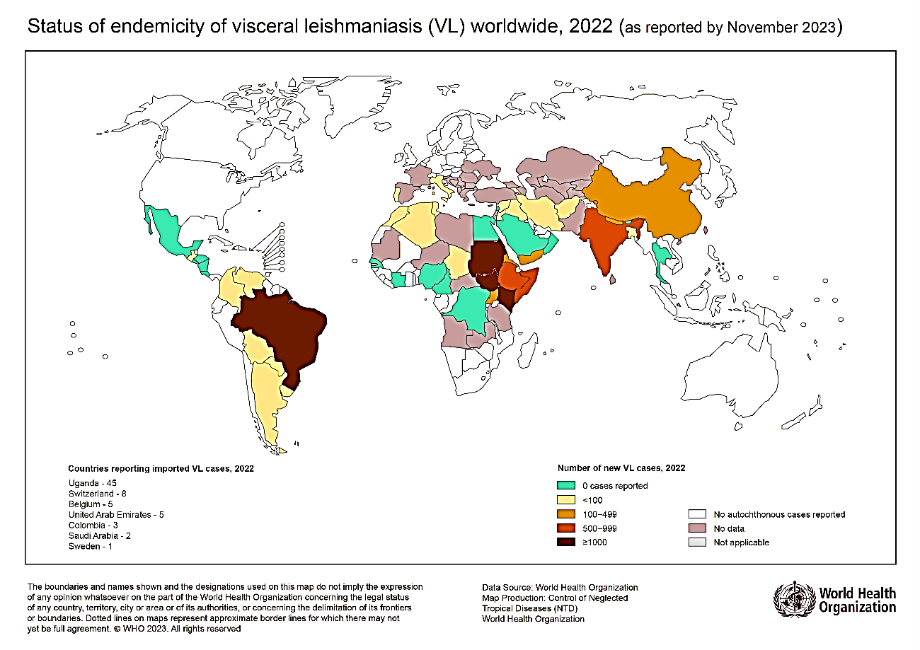
Source: World Health Organization (WHO): https://www.who.int/images/default-source/maps/leishmaniasis_vl_2022.png?sfvrsn=57d2c12f_3 7
An epidemiological shift has also been described since the 1980s, moving from a predominantly pediatric pattern (up to 70% of cases) to a current distribution where more than 75% of cases occur in adults, with a high proportion (50–60%) of HIV coinfection. In these groups, the estimated prevalence ranges from 2% to 3%, particularly in immunocompromised patients.⁵
International medical cooperation, including that carried out by Cuba in African countries, necessitates strengthening the capabilities of epidemiological surveillance, timely diagnosis, and clinical management of emerging and re-emerging diseases. It also involves exposure to nonendemic pathogens for healthcare personnel without prior immunity, which increases the complexity of the clinical approach.⁸⁻¹⁰
In this context, we present the following clinical case, which aims to describe the clinical, diagnostic, and therapeutic characteristics of a pediatric patient with confirmed visceral leishmaniasis treated in the Bioko Norte region of Equatorial Guinea, as well as to analyze the epidemiological implications and challenges in managing the disease in a setting with low endemicity and diagnostic limitations.
CLINICAL CASE
A 10-year-old pediatric patient who, for approximately 7 months, has had recurrent visits to emergency departments at various hospitals and clinics due to pallor of the skin and mucous membranes, with anemia confirmed by laboratory tests, anorexia, weight loss, and general malaise. His medical history included hospitalizations for hematologic disorders and treatment with recurrent blood transfusions over the past 4 months. He also had a Salmonella infection diagnosed via PDR, which was treated without complications. During the evaluation of anemia, gastroscopy was performed, revealing hyperplastic antral gastritis associated with Grade 2 enterogastric reflux, esophagitis, and a positive test for Helicobacter pylori, leading to the decision to admit the patient for treatment. Hemoglobin electrophoresis was deferred to rule out sickle cell disease because of a history of previous transfusions, and a fecal occult blood test was performed, which was positive. During a multidisciplinary case discussion involving gastroenterology, hematology, and pediatric specialists, gastrointestinal bleeding was suspected as the cause of the anemia, and treatment with intravenous (IV) omeprazole was initiated. Multiple hematological, biochemical, and serological tests were ordered to investigate anemia and establish a definitive diagnosis. One week after admission, with no change in his general condition or symptoms, he began to develop edema in the lower extremities and moderate free fluid in the abdominal cavity extending toward the hepatorenal and perisplenic areas, as detected by abdominal ultrasound. Additionally, a laminar pericardial effusion toward the left wall was identified.
Extraction and cytological and microbiological examination of the free abdominal fluid were recommended. The results revealed abundant mesothelial cells and polymorphonuclear lymphocytes. A positive result was confirmed by the GenXpert MTB/RIF nucleic acid amplification test. Extrapulmonary tuberculosis was diagnosed, and antituberculosis treatment was administered to the pediatric patient according to the regional regimen with Rifampicin/Isoniazid/Pyrazinamide/Ethambutol (RHZE).11 Discharge was proposed the following month, with clinical improvement evidenced by acceptable hematological values and a reduction in edema and ascites. Close follow-up by a pediatric clinic and a tuberculosis control program is recommended.
During the week following hospital discharge, the patient presented to the pediatric emergency department with progressive deterioration of the general condition, pallor, respiratory distress, a distended abdomen with positive signs of free fluid in the cavity (positive Tarral-Csonka maneuver), the presence of collateral circulation in the abdominal region, and generalized edema.
It was decided to admit the patient to the pediatric infectious diseases unit (Tisiologia) for comprehensive evaluation by specialists in pediatrics, hematology, and infectious diseases. Further bacteriological studies of the abdominal fluid were ordered; samples were collected and sent to a WHO-certified regional laboratory.
Physical examination. Moist mucous membranes with marked pallor. Respiratory system: decreased breath sounds in the right lung field with moderate intercostal and subcostal retractions. Respiratory rate (RR): 45 breaths per minute. The tissue oxygen saturation was 94%. Cardiovascular system: rhythmic, rapid heart sounds and the presence of a 1/6 systolic murmur at the left sternal border. Blood pressure (BP) was 95/70 mmHg. Heart rate (HR) 140 bpm. Abdomen: distended, with positive “Ola” and tarral signs and hepatosplenomegaly. Subcutaneous tissue: Generalized infiltrates present, with marked edema in the lower extremities. Central nervous system: conscious, oriented, with appropriate responses to questioning and preserved reflexes.
Laboratory findings. A complete blood test on admission revealed a hemoglobin concentration of 4 g/dL, a white blood cell count of 10 × 10³, a predominance of neutrophils, and a platelet count of 250 × 10³. Peripheral smears revealed red blood cells of variable size (anisocytosis), normal white blood cells, and normal platelets in terms of number and function. Biochemistry revealed the following: an albumin concentration of 1.58 g/dL, a creatinine concentration of 0.30 mg/dL, a total protein concentration of 0.9 g/dL, a ferritin concentration of 6.83 mg/mL, a GPT concentration of 15 U/L, a GOT concentration of 21.7 U/L, and a serum iron concentration of 54 μmol/L. Serology: negative for HBV, HCV, and HIV
Treatment. The patient was admitted while continuing RHZE treatment during the second month of administration for a weight of 24.5 kg, along with other pharmacological measures such as nutritional supplements and diuretic therapy, as well as nonpharmacological measures for symptomatic and syndromic treatment.11 Oral administration of 4 RHZ tablets (75 mg of rifampicin/50 mg of isoniazid/150 mg of pyrazinamide) diluted in 40 ml of water daily. Ethambutol (100 mg) 4 tablets diluted in water were orally administered separately from the daily RHZ administration. During the second week of treatment, with no significant clinical improvement, the results from the abdominal fluid analysis revealed a positive PCR result for Leishmania spp.
Multidisciplinary discussions were held among specialists in infectious diseases, pediatrics, and hematology to evaluate therapeutic options. It was decided to discontinue antituberculosis treatment and nutritional supplements, continue diuretic therapy, and initiate antiparasitic treatment with available medications. It was agreed to use liposomal amphotericin B at a dosage of 4 mg/kg daily, divided into 10 intermittent doses (days 1–5, with doses continuing on days 10, 17, 24, 31, and 38).4
Day 1 of treatment. Pre- and posttreatment hydration with 250 mL of 0.9% sodium chloride IV over two hours. Liposomal amphotericin B (1 mg/kg) was administered as a 30-minute tolerance test. Liposomal amphotericin B (3 mg/kg) was administered as a 2-hour infusion. Posttreatment flushing of the venous access with 5% glucose solution.
Days 4, 5, 10, 17, 24, 31, and 38 of treatment. Pre- and posttreatment hydration with 250 ml of 0.9% sodium chloride IV over two hours. Pretreatment (antipyretics, steroids). Liposomal amphotericin B (4 mg/kg) was administered as a 2-hour infusion. Other general interventions, such as daily parenteral hydration, continuous monitoring, infusion control, and treatment of adverse effects of antiparasitic therapy, are indicated, including antiemetics (nausea, vomiting), antipyretics (fever), and analgesics (myalgia).
The patient showed clinical improvement at 7 days, with resolution of general symptoms and edema. At 10 days, abdominal fluid was significantly reduced, as quantified by abdominal ultrasound. It was decided to extend the hospital stay until completion of treatment and periodic monitoring of liver and kidney function. The patient was discharged 40 days after the last admission. Follow-up was recommended, with readmissions every 3 months for 2 years for clinical evaluation, hematological tests, and relapse prevention. At the time of publication, the first periodic evaluation period had not yet been completed. On the basis of clinical criteria and laboratory findings, a diagnosis of visceral leishmaniasis was made.
DISCUSSION
The diagnosis of visceral leishmaniasis (VL) is characterized by a prolonged incubation period, which can range from 2 to 8 months, during which time the patient may remain asymptomatic while the hematological phase of the disease progresses.⁴ In the early stages, the clinical presentation is often nonspecific and may mimic other infectious conditions with high prevalence in endemic regions.⁴,⁸,¹⁰. In settings such as Central Africa, the differential diagnosis is particularly complex because of the coexistence of diseases such as malaria and salmonellosis, which share general clinical manifestations. In this scenario, the interpretation of rapid diagnostic tests (RDTs) may be limited by the population’s high prior exposure to various pathogens, which increases the likelihood of false-positive results.¹⁰ Furthermore, the possibility of coinfection between prevalent and neglected diseases should not be ruled out, which adds complexity to the clinical approach.¹⁰,¹² In the case presented, a previous positive RDT result for salmonellosis was documented, with an initially favorable clinical course. A diagnosis of tuberculosis was subsequently established using automated molecular tests with high sensitivity and specificity. However, the sample used did not correspond to the type recommended by the manufacturer of the diagnostic method, which limits the validity of the results.¹³ This aspect highlights the importance of proper selection of biological samples in settings with limited diagnostic resources. With respect to the LV, the parasite load in peripheral blood is usually low, which reduces the sensitivity of tests performed on this type of sample. In contrast, the examination of bone marrow or reticuloendothelial tissues (spleen, liver) offers greater diagnostic yield.¹⁴ In this context, performing a bone marrow aspiration would have allowed for the identification of amastigotes, contributing to diagnostic confirmation. Parasitic infiltration in these tissues also explains the observed hematological abnormalities, including anemia and cytopenias.
With respect to therapeutic management, some studies suggest that in the presence of coinfection, treatment of disease with greater clinical severity should be prioritized, with the goal of reducing complications and mortality.⁴ However, recent meta-analyses point to methodological limitations in the available studies, preventing the establishment of conclusive recommendations in this context.⁴,¹⁵ In the present case, the antituberculosis treatment initiated prior to the last admission failed to reverse clinical deterioration or the cardinal symptoms, which prompted a reconsideration of the diagnosis. The persistence of the clinical picture, along with the unfavorable course, led to the prioritization of specific treatments for LV, with a favorable clinical response observed after the initiation of antiparasitic therapy. This outcome retrospectively supports the accuracy of the definitive diagnosis.
According to the World Health Organization guidelines, there are various therapeutic options, including combination regimens.⁴ However, liposomal amphotericin B remains the treatment of choice because of its efficacy and safety profile, which have been extensively documented since its introduction in the 1990s.14,15,16 In this case, initial monotherapy was chosen, considering the patient’s clinical condition and the need for close monitoring during the initial phase of treatment. Measures were also implemented for the prevention and management of adverse events, with adequate tolerance and a minimal incidence of complications.
This case highlights the importance of strengthening epidemiological surveillance systems for neglected tropical diseases and training healthcare personnel in high-risk settings. Early clinical suspicion, together with access to appropriate and timely diagnostic methods, is essential for improving diagnostic accuracy in settings with a high burden of infectious diseases. However, it is necessary to consider the structural limitations and costs associated with these technologies, especially in resource-limited regions.
Overall, this case highlights the importance of integrating the epidemiological context, clinical course, and critical interpretation of diagnostic tests to guide the management of visceral leishmaniasis, particularly in nonendemic areas or those with low reporting rates, where diagnostic delay can significantly impact patient prognosis.
CONCLUSIONS
Visceral leishmaniasis is a neglected tropical disease of particular importance in settings where international medical cooperation takes place. The high prevalence of other infections with similar clinical manifestations, combined with acquired immunity in the population, contributes to initial misdiagnosis and delays the timely identification of this condition if an adequate clinical–epidemiological analysis is not performed. In this regard, integrating clinical suspicion with the rational use of diagnostic methods and the correct interpretation of results is essential for establishing an accurate diagnosis. Early implementation of specific therapeutic measures has a direct effect on the patient’s clinical course and prognosis. This case reinforces the need to maintain a broad and dynamic diagnostic perspective in complex settings, where neglected diseases may go unnoticed, reminding us that clinical success depends not only on available resources but also on the ability to continually question, integrate, and reevaluate clinical information.
References
- 1 Mañes Jiménez Y, Pedrón Marzal GM. Análisis de 37 casos de leishmaniasis en niños, diagnosticados en una comarca de Valencia, España. Rev Pediatr Aten Primaria [Internet]. 2021 [citado 1 julio 2025];23: 33-4. Disponible en: https://scielo.isciii.es/pdf/pap/v23n89/1139-7632-pap-89-23-33.pdf
- 2 Ministerio de Salud Pública y Bienestar Social Paraguay ¿Por qué lleva el nombre “Leishmaniasis” esta enfermedad? Departamento de Portales Web [Internet]. Mayo 2018 [citado 1 julio 2025]. Disponible en: https://www.mspbs.gov.py/portal/14821/iquestpor-que-lleva-el-nombre-leishmaniasis-esta-enfermedad.html
- 3 https://www.who.int/es/news-room/fact-sheets/detail/leishmaniasis
- 4 World Health Organization. WHO guidelines for the treatment of visceral leishmaniasis in HIV co-infected patients in East Africa and South-East Asia. World Health Organization [Internet]. 2022 [citado 1 julio 2025]. Disponible en: https://iris.who.int/server/api/core/bitstreams/d507ee8e-f280-4614-ac11-99cebf861579/content
- 5 Burza S Croft SL Boelaert M. Leishmaniasis. Lancet [Internet]. 2018 [citado 30 junio 2025];392: 951–70. Disponible en: https://doi.org/10.1016/S0140-6736(18)31204-2
- 6 La leishmaniasis visceral. https://www.elsevier.es/es-revista-medicina-integral-63-articulo-la-leishmaniasis-visceral-12967
- 7 World Health Organization (WHO). Status of endemicity of visceral leishmaniasis (VL) worldwide 2022. WHO [Internet]. 2023 [citado 30 junio 2025]. Disponible en: https://www.who.int/images/default-source/maps/leishmaniasis_vl_2022.png?sfvrsn=57d2c12f_3
- 8 Momeñe-Rojas NA, Pereda-Barroeta J, Villamandos-Tellaeche E, Díaz-Fernández M, Franco-Vicario R. Leishmaniasis visceral en paciente inmunocompetente. Rev Esp Casos Clin Med Intern. 2023; 8(2): 77-79. doi: https://doi.org/10.32818/reccmi.a8n2a6
- 9 Silva-Valido Jorge Alberto, Denis-de Armas Reinaldo, Franco-Bonal Arlene, Khanyile Victor, Mashiane Phuti. Experiencia de la Brigada Médica Cubana “Henry Reeve” en el enfrentamiento a la COVID-19 en Sudáfrica. Rev Inf Cient. [Internet]. 2021 [citado 30 junio 2025] ; 100( 6 ): Disponible en: http://scielo.sld.cu/scielo.php?script=sci_arttext&pid=S1028-99332021000600010&lng=es
- 10 Páez Arguelles O, Carrera Gonzalez E, Mojena EP, Auza Santivañez JC, Berty Gutiérrez H, Quisbert Vasquez HT, et al. Human African trypanosomiasis (sleeping sickness): a pediatric case report. South Health and Policy [Internet]. 2025 [citado 10 septiembre 2025]; 4:347. Disponible en: https://doi.org/10.56294/shp2025347
- 11 Organización Panamericana de la Salud. Manual operativo de la OMS sobre la tuberculosis. Módulo 5: Manejo de la tuberculosis en la población infantil y adolescente. OPS [Internet]; 2023 [citado 1 julio 2025]. Disponible en: https://doi.org/10.37774/9789275326930
- 12 Reyes-Baque JM, Loor-Solórzano MA, Moreira-Loor CS. Malaria: respuesta inmune, sensibilidad y especificidad de las pruebas rápidas. MQR Investigar [Internet]. 2024 [citado 25 octubre 2025];8(1):2617-32. Disponible en: https://www.investigarmqr.org/ojs/index.php/mqr/article/view/1017
- 13 Global Laboratorio Iniciative advancing TB diagnosis. Módulo 11: Guía clínica del Xpert MTB/RIF. Organización Panamericana de la Salud [Internet]. 2016 [citado 25 octubre 2025]. Disponible en: https://www.paho.org/sites/default/files/2016-cha-genexpert-mod-11.pdf
- 14 Verrest L, Kip AE, Musa AM, et al. Blood Parasite Load as an Early Marker to Predict Treatment Response in Visceral Leishmaniasis in Eastern Africa. ClinInfect Dis [Internet]. 2021 [citado 25 octubre 2025]; 73:775. Disponible en: https://doi.org/10.1093/cid/ciab124
- 15 Kotepui KU, Masangkay FR, De Jesus Milanez G, Kotepui M. Prevalence and outcomes of malaria as co-infection among patients with human African trypanosomiasis: a systematic review and meta-analysis. Sci Rep [Internet]. 2021[citado 30 junio 2025] ;11(1):23777. Disponible en: https://doi.org/10.1038/s41598-021-03295-8
Declarations
Funding
The authors received no funding for this study.
Conflict of interest
The authors declare that there are no conflicts of interest.
Authorship contributions
Conceptualization: Osmel Páez Arguelles, Elier Carrera González.
Data curation: Osmel Páez Arguelles, Elier Carrera González, Jhossmar Cristians Auza Santivañez, Neylin Carpio Negrin.
Formal analysis: Elier Carrera González and Neylin Carpio Negrin.
Research: Osmel Páez Arguelles, Elier Carrera González, Jhossmar Cristians Auza Santivañez, Neylin Carpio Negrin, Euliber Palomino Mojena
Methodology: Osmel Páez Arguelles, Elier Carrera González, Jhossmar Cristians Auza Santivañez,
Project management: Elier Carrera González.
Supervision: Jhossmar Cristians Auza Santivañez.
Visualization: Euliber Palomino Mojena, Neylin Carpio Negrin.
Drafting – original draft: Osmel Páez Arguelles, Elier Carrera Gonzalez, Euliber Palomino Mojena, Neylin Carpio Negrin, Jhossmar Cristians Auza-Santivañez, Freddy Ednildon Bautista-Vanegas, Henrry Temis Quisbert Vasquez, and Isaura Santander Oberson.
Writing–review and editing: Osmel Páez Arguelles, Elier Carrera Gonzalez, Euliber Palomino Mojena, Neylin Carpio Negrin, Jhossmar Cristians Auza-Santivañez, Freddy Ednildon Bautista-Vanegas, Henrry Temis Quisbert Vasquez, and Isaura Santander Oberson.