

Abstract
Inflammatory myofibroblastic tumor (IMT) is a distinctive neoplasm with limited metastatic potential. It is a rare disease in pediatrics, generally benign, although with possible progression to malignancy. We present the case of a 6-year-old female patient referred from a second-level hospital due to a clinical picture of large abdominal distension and a palpable abdominal mass. On admission, the patient presented slightly pale, hydrated mucous membranes, complained of abdominal discomfort, a distended abdomen, tender to superficial and deep palpation, with no peritoneal irritation. A giant abdominal mass was palpated occupying all quadrants of the abdomen, of hard consistency, tender to deep palpation, and hypoactive air sounds (AAR). An abdominal ultrasound was performed, which reported a retroperitoneal tumor lesion. Based on this finding, an abdominal and pelvic computed tomography (CT) scan with contrast was indicated, with the finding of a heterogeneous retroperitoneal tumor lesion likely related to neuroblastoma. Based on all the findings, the patient was admitted to the operating room for exploratory laparotomy and open biopsy. Transoperatively, a giant abdominal mass was revealed. Histopathological findings were consistent with inflammatory myofibroblastic tumor. The diagnosis was confirmed with immunohistochemistry. A comprehensive multidisciplinary evaluation was performed, and surgical intervention was initiated, with favorable outcomes. From a therapeutic perspective, complete surgical resection remains the mainstay of treatment, as it was in our clinical case, especially in localized and resectable lesions, as it is associated with a lower risk of recurrence. This clinical case highlights the inherent complexity of addressing IMT in pediatric patients, reinforcing the need for close collaboration between pediatricians, surgeons, imaging specialists, pathologists, and oncologists. Furthermore, due to the rarity of the disease in the pediatric population, further research and collective experience are needed to improve our understanding of IMT and refine treatment strategies for these cases.
Keywords
Inflammatory Myofibroblastic Tumor, Tumor Markers, Inflammatory Pseudotumor, Surgery
INTRODUCTION
Inflammatory myofibroblastic tumor (IMT) is a distinctive neoplasm with limited metastatic potential, characterized by the proliferation of fibroblastic and myofibroblastic spindle cells, accompanied by a dense inflammatory infiltrate composed mainly of plasma cells, lymphocytes, and eosinophils.(1) It is a rare disease in children, generally benign, although it can progress to malignancy. ITMT is a rare mesenchymal neoplasm, accounting for less than 1 % of all soft tissue tumors. Its exact prevalence is difficult to determine due to its rarity, histological heterogeneity, and historical misclassification as a reactive or inflammatory process. It predominantly affects children and young adults, but occurs across a wide age spectrum.(2,3) There is a slight female predominance; however, the location of the tumor may affect both the age of onset and gender distribution. SITs can occur in the abdominal cavity (75 % of cases), particularly in the mesentery, greater omentum, and retroperitoneal space; as well as in the head and neck, lungs, urinary bladder, central nervous system, and female genital tract. They mainly affect children and young adults.(4,5) Gastrointestinal IMTs predominantly affect the small intestine and colon, followed by the stomach. Less frequently, they affect the esophagus, appendix, pancreas, and liver.(6)
Accurate pathological classification and a comprehensive understanding of the molecular pathogenesis of MIT are crucial for improving treatment and prognosis in patients. 7Currently, MITs are classified as rare intermediate-grade neoplasms, which have a high recurrence rate after excision and exhibit low metastatic potential. However, diagnosis and treatment remain complex due to their considerable histological and molecular heterogeneity.(8,9,10,11,12) We present the case of a pediatric patient with this complex pathology, perform a comprehensive and multidisciplinary assessment, and take surgical action with favorable results.
CASE REPORT
A 6-year-old female patient from the Yungas region of La Paz, Bolivia, was referred from a secondary care hospital with a clinical picture of severe abdominal distension and a palpable abdominal mass. In addition, the family re t reported that she suffers from constipation. On physical examination upon admission, the positive findings include slightly pale, hydrated mucous membranes, abdominal discomfort, distended abdomen, tenderness on superficial and deep palpation, no peritoneal irritation, and a giant abdominal mass measuring approximately 15 x 10 cm in diameter, occupying all quadrants of the abdomen, hard in consistency, and painful on deep palpation. Hypoactive bowel sounds (HBS).
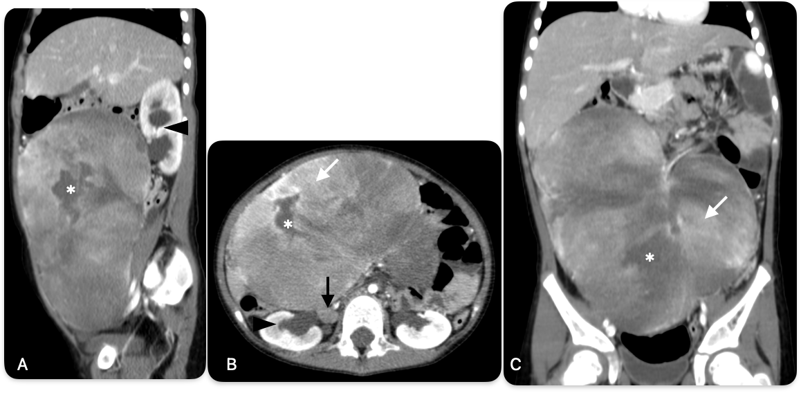
An abdominal ultrasound was performed, which reported a retroperitoneal tumor lesion causing a high diaphragm due to cranial displacement of peritoneal contents, measuring 17,2 x 15,9 x 10 cm with a volume of 1436 ml, suggesting a probable neuroblastoma, without ruling out other pathologies, high diaphragm due to cranial displacement of peritoneal contents. Given this finding, a contrast-enhanced computed tomography (CT) scan of the abdomen and pelvis was indicated (figure 1), revealing a heterogeneous retroperitoneal tumor lesion likely related to neuroblastoma, measuring approximately 19,4 x 9,8 x 16,4 cm (L x AP x T) with a volume of 1621 ml, a cluster of retroperitoneal adenopathies related to secondary primary disease, and grade III-IV right ureterohydronephrosis secondary to extrinsic compression of a retroperitoneal lesion.
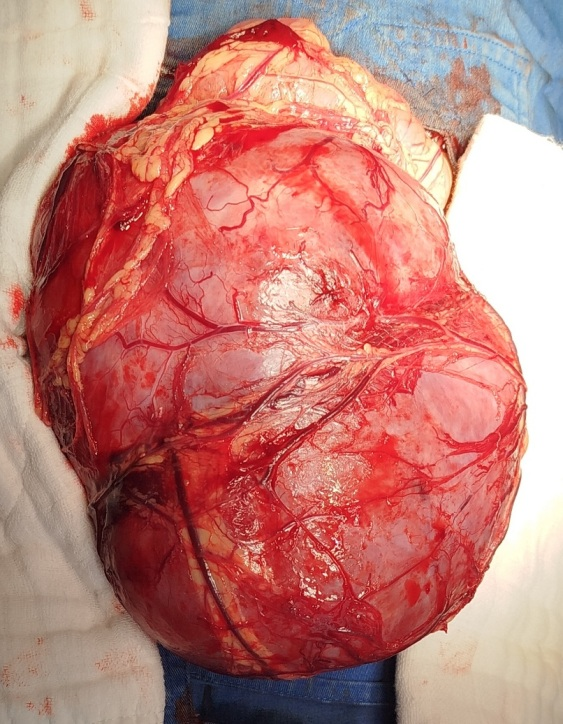
Based on all findings, it was decided to admit the patient to the operating room for exploratory laparotomy and open biopsy. Intraoperatively, a giant abdominal mass was found (figure 2), with both cystic and solid components and neovascularization. An incisional biopsy was performed, revealing bleeding in the napa and abundant bleeding at the biopsy site. Hemostatic maneuvers were performed to contain the bleeding. When active bleeding was observed, total resection of the tumor was performed, the incision was extended to supra and infraumbilical areas, and total resection of the tumor was performed, in addition to partial resection of the greater omentum involved in the mass, supported by the use of a harmonic scalpel during surgery.
The histopathological report shows in its microscopic description: The histological sections show a hypocellular neoplasm consisting of parallel spindle-shaped myofibroblasts with elongated nuclei, vesicular chromatin, and insinuated nucleoli; some cells have elongated nuclei with macronucleoli or chromatin, arranged in abundant myxoid stroma with the presence of inflammatory cells consisting of lymphocytes, plasmocytes, some neutrophils, and histiocytes, few mitotic figures, areas of hemorrhage and vascular congestion, peripherally surrounded by a thin fibroconnective capsule. Conclusion: Histopathological findings consistent with inflammatory myofibroblastic tumor; diagnosis confirmed by immunohistochemical study.
DISCUSSION
Inflammatory myofibroblastic tumor (IMT) is a rare and uncommon condition classified as an inflammatory pseudotumor. It most commonly forms in two types of tissue: the mucous membranes and the mesentery. It can occur in any age group, but is most prevalent in children and adolescents. The clinical symptoms of ITM are usually specific to each organ. Computed tomography (CT) is the most reliable diagnostic method. In our case, a CT scan was performed before the operation on the 6-year-old girl, which helped guide the formulation of the surgical plan. Laboratory test results vary considerably. The final diagnosis still depends on the pathological and immunohistochemical results. The clinical presentation is related to the location of the tumor. In most cases, it is asymptomatic, but a percentage of cases may present with general manifestations such as asthenia, anorexia, weight loss, fever, and microcytic and hypochromic anemia. Diagnosis of the mesenteric variety is difficult due to its clinical and imaging presentation, as well as its variable characteristics, as demonstrated in the present clinical case, where the differential diagnosis and, above all, imaging revealed other types of retroperitoneal tumors. Even the transsurgical findings showed a large abdominal mass with cystic and solid components, as well as neovascularization suggestive of a neoplasm. The new technological impact in imaging with vascular and non-vascular interventional techniques in medicine provides an adjunct to the treatment and management of this type of lesion with embolization of the main polar arteries that feed the tumor before its total resection.New magnetic resonance imaging sequences and the use of gadolinium contrast agents provide new findings for diagnostic imaging and complement the study of benignity and malignancy in this type of lesion for its proper management.
The differential diagnosis of MIT is made with benign lesions such as cystic lymphangiomas, giant cell granuloma, solitary fibrous tumor, myxepithelioma, myxofibroma, and malignant tumors such as neuroblastoma, teratomas, lymphomas, sarcoma, and rhabdomyosarcomas. Surgery remains the treatment of choice; however, Zhao et al. presented two cases of unresectable intra-abdominal inflammatory myofibroblastic tumors that experienced spontaneous regression without surgical treatment. Total surgical resection of the tumor is the surgical treatment of choice, since complete surgical resection is curative; when the tumor is completely resected, the mass effect in the abdominal cavity disappears, and all symptoms and abnormalities disappear. Recurrences have been reported when resection is incomplete.
The literature mentions that in none of the cases reviewed was inflammatory myofibroblastic tumor diagnosed, either before or during surgery. The definitive diagnosis was made by pathological anatomy through histopathological and immunohistochemical studies.
CONCLUSIONS
Inflammatory myofibroblastic tumor (IMT) is a rare neoplastic entity in children, with clinical, histopathological, and molecular characteristics that make it a significant diagnostic and therapeutic challenge. Its intermediate nature, with low metastatic potential but a high rate of local recurrence, requires comprehensive evaluation and a multidisciplinary approach for proper characterization and management. Accurate diagnosis of IM requires not only detailed morphological evaluation but also the systematic use of immunohistochemical techniques and molecular studies, especially those aimed at detecting genetic rearrangements such as those of the ALK gene, which have both prognostic and therapeutic implications, which is limited in our context. From a therapeutic standpoint, complete surgical resection remains the mainstay of treatment, as it was in our clinical case, especially in localized and resectable lesions, as it is associated with a lower risk of recurrence. This clinical case highlights the inherent complexity of the approach to MTI in pediatric patients, reinforcing the need for close collaboration between pediatricians, surgeons, imaging specialists, pathologists, and oncologists. Furthermore, due to the rarity of the disease in the pediatric population, more research and collective experience are needed to improve our knowledge of MTI and refine treatment strategies for these cases.
References
- 1 Yamamoto H. Inlammatory myoibroblastic tumour. En: The WHO Editorial Board E, editor. WHO classification of tumours of soft tissue and bone tumours, 5th ed. 5a ed. Lyon, France: IARC; 2020. p. 109–11.
- 2 Coffin CM, Hornick JL, Fletcher CDM. Inflammatory myofibroblastic tumor: comparison of clinicopathologic, histologic, and immunohistochemical features including ALK expression in atypical and aggressive cases. Am J Surg Pathol. 2007;31(4):509–20. Disponible en: http://dx.doi.org/10.1097/01.pas.0000213393.57322.c7
- 3 Coffin CM, Watterson J, Priest JR, Dehner LP. Extrapulmonary inflammatory myofibroblastic tumor (inflammatory pseudotumor). A clinicopathologic and immunohistochemical study of 84 cases. Am J Surg Pathol. 1995;19(8):859–72. Disponible en: http://dx.doi.org/10.1097/00000478-199508000-00001
- 4 Cassarino DS, Gardner J, Lucas D, Quick CM, Stallings-Archer K. Diagnostic pathology: Soft tissue tumors third ed. 3a ed. Vols. 88–99. Philadelphia: Elsevier - Health Sciences Division; 2019. 114-791
- 5 Mocellin S. Soft tissue tumors: A practical and comprehensive guide to sarcomas and benign neoplasms. Cham: Springer International Publishing; 2021.
- 6 Casanova M, Brennan B, Alaggio R, Kelsey A, Orbach D, van Noesel MM, et al. Inflammatory myofibroblastic tumor: The experience of the European pediatric Soft Tissue Sarcoma Study Group (EpSSG). Eur J Cancer. 2020; 127:123–9. Disponible en: http://dx.doi.org/10.1016/j.ejca.2019.12.021
- 7 Armstrong V, Khazeni K, Rosenberg A, Swain SK, Moller M. Inflammatory pseudotumor secondary to urachal cyst: A challenging clinical case report. Int J Surg Case Rep. 2020; 66:360–4. http://dx.doi.org/10.1016/j.ijscr.2019.12.029
- 8 Antonescu CR, Blay JY, Bovee JVGM, Bridge JA, Cunha IW, Dei Tos AP, Flanagan AM, Fletcher CDM, Flope AL, Hornick JL, Mertens F, Miettines M,Nielsen GP, Oda Y, Yoshida A. WHO classification of tumours of soft tissue and bone tumours, 5th ed. 5a ed. Vols. 2–4. Lyon, France: IARC; 2020.
- 9 Callizaya Macedo G, Auza-Santivañez JC, Vallejos Rejas DRE, Quispe Sarmiento RA, Flores Canaviri JJ, Suarez Laime LH. Giant multiloculated omental cyst in a pediatric patient. Case report and literature review. Multidisciplinar (Montevideo). 2024; 2:88. https://doi.org/10.62486/agmu202488
- 10 Coffin CM, Hornick JL, Fletcher CDM. Inflammatory myofibroblastic tumor: comparison of clinicopathologic, histologic, and immunohistochemical features including ALK expression in atypical and aggressive cases. Am J Surg Pathol. 2007; 31(4):509–20.
- 11 Aguirre Cruz B, Auza-Santivañez JC, Vallejos-Rejas DRE, Mendoza-Contreras E, Quispe-Medrano V, Salazar Ortega BF, et al. Usefulness of Ultrasound in the diagnosis of Septic Arthritis: about a case. AG Salud. 2025; 3:192. https://doi.org/10.62486/agsalud2025192
- 12 Quisbert Vasquez HT, Auza-Santivañez JC, Moscoso Zurita G, Suarez Laime LH, Apaza Huanca B, Callizaya Macedo G. Choledochal cyst in a pediatric patient with Down syndrome, a diagnostic challenge. SCT Proceedings in Interdisciplinary Insights and Innovations. 2025; 3:379. https://doi.org/10.56294/piii2025379
Declarations
Funding
The authors did not receive funding for the application of this study.
Conflict of interest
The authors declare that there is no conflict of interest.
Authorship contributions
Conceptualization: Giovanni Callizaya Macedo, Jhossmar Cristians Auza-Santivañez
Formal analysis: Giovanni Callizaya Macedo
Research: Freddy Ednildon Bautista-Vanegas, Jhossmar Cristians Auza-Santivañez
Methodology: Giovanni Callizaya Macedo
Project management: Ingrid Neysa Cabezas-Soliz, Giovanni Callizaya Macedo
Supervision: Giovanni Callizaya Macedo
Visualization: Ritdber Alfredo Quispe Sarmiento
Writing – original draft: Giovanni Callizaya Macedo, Jhossmar Cristians Auza-Santivañez, Ingrid Neysa Cabezas-Soliz, Martin Felix Fernando Alarcón Caba, María Angela Murillo Alcázar, Mayra Estephany Atila Lujá, Ritdber Alfredo Quispe Sarmiento, Freddy Ednildon Bautista-Vanegas
Editing – review and editing: Giovanni Callizaya Macedo, Jhossmar Cristians Auza-Santivañez, Ingrid Neysa Cabezas-Soliz, Martin Felix Fernando Alarcón Caba, María Angela Murillo Alcázar, Mayra Estephany Atila Lujá, Ritdber Alfredo Quispe Sarmiento, Freddy Ednildon Bautista-Vanegas