
Abstract
Preoperative radiotherapy plays a crucial role in the treatment of rectal cancer. It is the only method proven to significantly reduce local recurrence rates. Radiotherapy contributes to improved treatment outcomes through three primary mechanisms: (1) downstaging the tumor to facilitate surgical resection, (2) reducing the risk of local recurrence by eradicating microscopic tumor foci in the operative field, and (3) increasing the likelihood of sphincter-preserving surgery, particularly in cases involving invasion of the levator ani or external anal sphincter muscles Method: in this study, we analyzed 289 patients with rectal cancer, 80 (27,6 %) of whom received preoperative radiotherapy. Of these, 30 patients (37,5 %) underwent short-course radiotherapy and 50 (62,5 %) underwent long-course radiotherapy. Among the long-course group, 22 patients (44 %) were in the laparoscopic surgery group and 28 (56 %) in the open surgery group. Long-course radiotherapy was delivered at 2 Gy per session over four weeks, while short-course radiotherapy consisted of 5 Gy per session over five days. Long-course results were assessed eight weeks post-radiotherapy. Discussion: short-course radiotherapy was primarily administered in cases with suspected mesorectal lymph node metastases, followed by total mesorectal excision (TME) surgery within the subsequent week. In patients who received long-course radiotherapy, three distinct response patterns were observed: complete radiosensitivity, partial radiosensitivity, and radioresistance. In cases of complete radiosensitivity, the tumor underwent total regression; in partially radiosensitive cases, tumor size was reduced but not completely eliminated. In radioresistant cases, no significant change in tumor size was observed following radiotherapy. To quantitatively assess these effects, tumor regression rates were evaluated. Results: following long-course radiotherapy, among patients in the laparoscopic group, 4 (18,2 %) demonstrated complete radiosensitivity, 15 (68,2 %) exhibited partial radiosensitivity, and 3 (13,6 %) were radioresistant. In the open surgery group, 5 (17,9 %) patients showed complete radiosensitivity, 19 (67,9 %) had partial radiosensitivity, and 4 (14,3 %) were radioresistant (p = 0,998). Regarding tumor regression grading, in the laparoscopic group, Grade 1 regression was 19,1 ± 5,7 %, Grade 2 was 51,1 ± 7,3 %, Grade 3 was 17,0 ± 5,5 %, and Grade 4 was 12,8 ± 4,9 %. In the open surgery group, the respective values were 23,2 ± 5,6 %, 44,6 ± 6,6 %, 17,9 ± 5,1 %, and 14,3 ± 4,7 %. Conclusion: long-course radiotherapy demonstrated efficacy in reducing tumor size (including instances of complete tumor regression), minimizing local recurrence, and increasing the feasibility of surgical intervention. No statistically significant differences were observed between the laparoscopic and open surgery groups in terms of radiosensitivity or tumor regression rates (p > 0,05). Notably, Grade 2 regression was the most prevalent outcome, observed in 51,1 ± 7,3 % of laparoscopic cases and 44,6 ± 6,6 % of open surgery cases.
Keywords
Radiotherapy, Short-Course Radiotherapy, Long-Course Radiotherapy, Rectal Cancer, Circumferential Resection Margin
INTRODUCTION
Radical surgical resection remains the cornerstone of treatment for malignant rectal tumors.(1,2) However, surgery alone is often associated with a high risk of local recurrence and, in certain cases, may be technically unfeasible due to tumor extent or anatomical constraints.(3,4,5) Furthermore, management of local recurrence is frequently limited and poses significant therapeutic challenges. Among available modalities, preoperative radiotherapy has been shown to be the most effective strategy for reducing local recurrence rates.(3,6,7,8) Currently, neoadjuvant radiochemotherapy followed by total mesorectal excision (TME) is considered the standard of care for locally advanced rectal cancer and is widely implemented in clinical practice worldwide.(1,2,4) The addition of radiochemotherapy to surgical treatment serves multiple purposes: it reduces the risk of local recurrence, facilitates tumor downstaging to improve resectability, and enhances the likelihood of sphincter preservation in low-lying tumors.(3,4,5) This study aims to evaluate the role of neoadjuvant radiochemotherapy in decreasing tumor size and minimizing recurrence in patients with rectal cancer.(9,10)
METHODS
The study included 80 out of 103 patients diagnosed with rectal adenocarcinoma between 2016 and 2021. Among these patients, 35 were female and 45 were male, with an age range of 19 to 78 years. Tumor localization was categorized as lower (0–6 cm), middle (7–12 cm), and upper rectum (>12 cm) based on the distance from the anal verge (table 1).
Distance from Anal Verge | Laparoscopic Group (n=47) | Laparoscopic Group (n=47) | Open Group (n=56) | Open Group (n=56) |
|---|---|---|---|---|
Distance from Anal Verge | n | % | n | % |
0-6 sm | 13 | 27,7 | 16 | 28,6 |
7-12 sm | 18 | 38,3 | 22 | 39,3 |
>12 sm | 16 | 34,0 | 18 | 32,1 |
χ2; p | χ2=0,042; p = 0,979 | χ2=0,042; p = 0,979 | χ2=0,042; p = 0,979 | χ2=0,042; p = 0,979 |
Note: the p-value represents the statistical significance calculated using Pearson’s polychoric correlation coefficient based on the analyzed trait indicators. | Note: the p-value represents the statistical significance calculated using Pearson’s polychoric correlation coefficient based on the analyzed trait indicators. | Note: the p-value represents the statistical significance calculated using Pearson’s polychoric correlation coefficient based on the analyzed trait indicators. | Note: the p-value represents the statistical significance calculated using Pearson’s polychoric correlation coefficient based on the analyzed trait indicators. | Note: the p-value represents the statistical significance calculated using Pearson’s polychoric correlation coefficient based on the analyzed trait indicators. |
In the laparoscopic and open surgery groups, the T-stage distribution was as follows: T1 – 3 patients (6,4 %) vs 2 patients (3,6 %), T2 – 11 (23,4 %) vs 10 (17,9 %), T3 – 29 (61,7 %) vs 36 (64,3 %), and T4 – 4 (8,5 %) vs 8 (14,3 %) (p = 0,668).
Stage-wise distribution was: Stage I – 7 patients (14,9 %) vs 5 patients (8,9 %), Stage II – 12 (25,5 %) vs 16 (28,6 %), and Stage III – 28 (59,6 %) vs 35 (62,5 %) (p = 0,637). Given the clinical significance of advanced tumor invasion, we considered it appropriate to classify patients with T3 and T4 tumors into a separate subgroup for further analysis (table 2).
T (mm) | T (mm) | n | Laparoscopic Group n=33 | Laparoscopic Group n=33 | n | Open Group n=42 | Open Group n=42 |
|---|---|---|---|---|---|---|---|
T (mm) | T (mm) | n | n | % | n | n | % |
pT3 | pT3a (<1) | 29 | 6 | 18,2 | 36 | 7 | 16,7 |
pT3 | pT3b (1-5) | 29 | 6 | 18,2 | 36 | 8 | 19,1 |
pT3 | pT3c (5-15) | 29 | 8 | 24,2 | 36 | 11 | 26,3 |
pT3 | pT3d (>15) | 29 | 9 | 27,3 | 36 | 10 | 23,8 |
pT4 | pT4a (i.v.) | 4 | 1 | 3,0 | 8 | 3 | 7,2 |
pT4 | Pt4b (i.y.) | 4 | 3 | 9,1 | 8 | 5 | 11,9 |
Note: the depth of tumor spread within the mesorectum was categorized as follows: pT3a – invasion up to 1 mm, pT3b – invasion of 1–5 mm, pT3c – invasion of 5–15 mm, and pT3d – invasion greater than 15 mm | Note: the depth of tumor spread within the mesorectum was categorized as follows: pT3a – invasion up to 1 mm, pT3b – invasion of 1–5 mm, pT3c – invasion of 5–15 mm, and pT3d – invasion greater than 15 mm | Note: the depth of tumor spread within the mesorectum was categorized as follows: pT3a – invasion up to 1 mm, pT3b – invasion of 1–5 mm, pT3c – invasion of 5–15 mm, and pT3d – invasion greater than 15 mm | Note: the depth of tumor spread within the mesorectum was categorized as follows: pT3a – invasion up to 1 mm, pT3b – invasion of 1–5 mm, pT3c – invasion of 5–15 mm, and pT3d – invasion greater than 15 mm | Note: the depth of tumor spread within the mesorectum was categorized as follows: pT3a – invasion up to 1 mm, pT3b – invasion of 1–5 mm, pT3c – invasion of 5–15 mm, and pT3d – invasion greater than 15 mm | Note: the depth of tumor spread within the mesorectum was categorized as follows: pT3a – invasion up to 1 mm, pT3b – invasion of 1–5 mm, pT3c – invasion of 5–15 mm, and pT3d – invasion greater than 15 mm | Note: the depth of tumor spread within the mesorectum was categorized as follows: pT3a – invasion up to 1 mm, pT3b – invasion of 1–5 mm, pT3c – invasion of 5–15 mm, and pT3d – invasion greater than 15 mm | Note: the depth of tumor spread within the mesorectum was categorized as follows: pT3a – invasion up to 1 mm, pT3b – invasion of 1–5 mm, pT3c – invasion of 5–15 mm, and pT3d – invasion greater than 15 mm |
During the pathohistological evaluation of the surgical specimens, adenocarcinoma with varying degrees of differentiation was identified in the majority of cases (table 3). In both study groups, moderately differentiated adenocarcinoma was the most frequently observed subtype, occurring in 57,4 % of laparoscopic cases and 55,4 % of open surgery cases (p = 0,998). Mucinous adenocarcinoma and squamous cell carcinoma were the least commonly detected histological types, accounting for 2,1 % and 1,8 % of cases, respectively, in the laparoscopic and open surgery groups.
Morphological Type | Morphological Type | Laparoscopic Group n=47 | Laparoscopic Group n=47 | Open Group n=56 | Open Group n=56 |
|---|---|---|---|---|---|
Morphological Type | Morphological Type | n | % | n | % |
Adenocarcinoma | High-grade differentiation Moderate differentiation Low-grade differentiation | 13 27 5 | 27,7 57,4 10,6 | 16 31 7 | 28,6 55,4 12,5 |
Mucinous carcinoma (colloid) | Mucinous carcinoma (colloid) | 1 | 2,1 | 1 | 1,8 |
Squamous cell carcinoma | Squamous cell carcinoma | 1 | 2,1 | 1 | 1,8 |
χ²; p-value | χ²; p-value | χ² = 0,134; p = 0,998 | χ² = 0,134; p = 0,998 | χ² = 0,134; p = 0,998 | χ² = 0,134; p = 0,998 |
Tumor staging was performed according to the TNM classification system. Prior to surgery, tumor location was identified as upper rectum (20 patients), middle rectum (36 patients), and lower rectum (24 patients). In the laparoscopic group, 35 patients received preoperative radiotherapy—13 patients received short-course radiotherapy, and 22 received long-course radiotherapy. In the open surgery group, 45 patients underwent radiotherapy—17 patients received short-course and 28 received long-course regimens.
Among patients with upper rectal tumors, in the laparoscopic group, 6 received short-course and 3 received long-course radiotherapy; in the open group, 7 received short-course and 4 received long-course radiotherapy. For middle rectal tumors, 8 patients in the laparoscopic group and 10 in the open group received short-course radiotherapy, while 8 and 10 patients, respectively, received long-course radiotherapy. For lower rectal tumors, no patients in either group received short-course radiotherapy; however, 11 patients in the laparoscopic group and 13 in the open group received long-course radiotherapy (table 4).
The long-course radiotherapy protocol consisted of 2 Gy fractions over 4 weeks, while the short-course protocol involved 5 Gy fractions administered over 5 consecutive days. The therapeutic response to long-course radiotherapy was assessed 8 weeks after completion of treatment.
Localization | TNM | Laparoscopic Group (n=35) | Laparoscopic Group (n=35) | Open Group (n=45) | Open Group (n=45) |
|---|---|---|---|---|---|
Localization | TNM | Short | Long | Short | Long |
High | T1-2 N1-2 M0 (within the mesorectum) | 1 | - | 2 | - |
High | T1-2N1-N2 M0 (within the mesorectum) | - | - | - | - |
High | T3N0 (SRS-) | - | - | - | - |
High | T3N1-2 (SRS-) (within the mesorectum) | 5 | - | 5 | - |
High | T3N1-2 (SRS-) (outside the mesorectum) | - | - | - | 1 |
High | T3 SRS+ | - | - | - | - |
High | T4NX | - | 3 | - | 3 |
Middle | T1-2N1-2 (within the mesorectum) | 2 | - | 2 | - |
Middle | T1-2N1-2 (outside the mesorectum) | - | - | - | 1 |
Middle | T3N0 (SRS-) | 3 | - | 6 | - |
Middle | T3N1-2 (SRS-) (within the mesorectum) | 2 | - | 2 | - |
Middle | T3N1-2 (SRS-) (outside the mesorectum) | - | 6 | - | 4 |
Middle | T3 SRS+ | - | 1 | - | 2 |
Middle | T4NX(0,1.2) | - | 1 | - | 3 |
Low | T1-2N1-2 (within the mesorectum) | - | 3 | - | 1 |
Low | T1-2N1-2 (outside the mesorectum) | - | 1 | - | - |
Low | T3N0 (SRS-) | - | 4 | - | 5 |
Low | T3N1-2 (SRS-) (within the mesorectum) | - | 1 | - | 3 |
Low | T3N1-2 (SRS-) (outside the mesorectum) | - | 1 | - | 2 |
Low | T3 SRS+ | - | 1 | - | 1 |
Low | T4NX | - | - | - | 2 |
Total | Total | 13 | 22 | 17 | 28 |
Of the patients who received radiotherapy, 13 were classified as stage T1–T2, 55 as stage T3, and 12 as stage T4. While the preoperative segmentation of the rectum into upper, middle, and lower thirds plays an important role in MRI-based evaluation, we believe that the anatomical position relative to the peritoneal reflection is particularly critical for accurately determining tumor location during surgery.
In this regard, when determining tumor location, we specifically recorded tumors located above and below the peritoneal reflection, as well as their distance from the peritoneal fold to the rectosigmoid junction. All patients included in the study (n = 80) underwent total mesorectal excision (TME) and double-lumen loop ileostomy. Patients with distant metastases were excluded from the study.
DISCUSSION
Based on both the literature and our clinical experience, we conclude that preoperative radiochemotherapy is recommended in all cases with lymph node-positive rectal cancer. In cases of T3N0M0 and T4N0M0 disease, particularly when abdominoperineal resection is planned, neoadjuvant therapy should be administered according to established clinical protocols.
For practical surgical classification, we divided the rectum anatomically into two main regions: (1) the supralevator part and (2) the infralevator part. The supralevator part was further subdivided into three segments: the upper, middle, and lower rectum. It is noteworthy that the middle and lower rectum are predominantly located below the peritoneal reflection, while the upper rectum lies above it. We consider inclusion of the peritoneal reflection as a reference point in rectal tumor classification to be clinically valuable, particularly for surgical planning and staging.
In this study, tumors arising from the sublevator (pelvic floor) region were not included. For preoperative radiochemotherapy, we applied both the short-course Swedish protocol and the long-course English protocol, tailored to the stage and anatomical location of the tumor.
All major colorectal cancer centers consider T3aN–T4 tumors as locally advanced. Neoadjuvant therapy is the standard recommendation for T3 and T4 tumors. If these tumors are resected without neoadjuvant therapy, adjuvant treatment is required postoperatively. Studies have demonstrated that preoperative radiotherapy is associated with lower rates of local recurrence and reduced radiation-related toxicity compared to postoperative treatment.
However, the optimal management of T3M0 tumors, particularly as assessed by MRI, remains controversial. Given the heterogeneity of T3 lesions, the prognosis is significantly poorer in T3c and T4 tumors. An important observation from our study is that among 17 patients clinically staged as T3N0, 4 patients (23,5 %) were found to have pathologically positive lymph nodes following surgery. This supports the recommendation for preoperative radiotherapy even in T3N0M0 tumors.
Additionally, 12 patients with T1–T2 tumors and confirmed nodal involvement also received preoperative radiotherapy. In 15 patients with suspected mesorectal fascia involvement, where the risk of a positive circumferential resection margin (CRM) was high, we administered long-course neoadjuvant chemoradiotherapy to reduce this risk.
In the neoadjuvant treatment setting, the short-course Swedish protocol (25 Gy in 5 fractions) was administered to 30 patients, while long-course chemoradiotherapy was given to 50 patients with locally advanced rectal tumors (T3N1–T4) and lower rectal tumors. The outcomes of patients who received long-course preoperative radiotherapy were evaluated based on changes in tumor size as observed on MRI and histopathological tumor regression.
The response to long-course radiotherapy was categorized into three groups: radiosensitive, partially radiosensitive, and radioresistant, as detailed in table 5.
Table 5. Tumor Tissue Response to Long-Course Radiotherapy in Both Laparoscopic and Open Surgery Patient Groups
Response Type | Laparoscopic Group (n=22) | Laparoscopic Group (n=22) | Open Group (n=28) | Open Group (n=28) |
|---|---|---|---|---|
Response Type | n | % | n | % |
Complete Response | 4 | 18,2 | 5 | 17,9 |
Partial Response | 15 | 68,2 | 19 | 67,9 |
Radioresistant | 3 | 13,6 | 4 | 14,3 |
χ2; p | χ2=0,005; p = 0,998 | χ2=0,005; p = 0,998 | χ2=0,005; p = 0,998 | χ2=0,005; p = 0,998 |
As a result of long-course radiotherapy, 4 patients (18,2 %) in the laparoscopic group exhibited complete radiosensitivity, 15 patients (68,2 %) had partial radiosensitivity, and 3 patients (13,6 %) were classified as radioresistant. In the open surgery group, 5 patients (17,9 %) had complete radiosensitivity, 19 patients (67,9 %) showed partial radiosensitivity, and 4 patients (14,3 %) were radioresistant (p = 0,998).
Among those who received long-course radiotherapy, 18,2 % of patients in the laparoscopic group and 17,9 % in the open group exhibited complete radiosensitivity. Standard total mesorectal excision (TME) was performed in all patients. Histopathological examination of the resected specimens revealed tumor regression in patients who underwent long radiotherapy, with the most significant regression observed in grade 2 (table 6).
In addition to these findings, other clinical parameters were comprehensively examined in patients who underwent total mesorectal excision and radiotherapy.
Table 6. Univariate Analysis of Factors Influencing the Outcome of Rectal Cancer Treatment
Indicator | Indicator | Laparoscopic Group | Open Group | p |
|---|---|---|---|---|
Indicator | Indicator | n (P±mp%) / M (min – max) | n (P±mp%) / M (min – max) | p |
TME Quality | High Quality | 23 (48,9±7,3 %) | 26 (46,4±6,7 %) | 0,952 |
TME Quality | Medium Quality | 15 (31,9±6,8 %) | 18 (32,1±6,2 %) | 0,952 |
TME Quality | Low Quality | 9 (19,1±5,7 %) | 12 (21,4±5,5 %) | 0,952 |
SRS Positivity | High 1/3 | 2/13 (15,4±10,0 %) | 2/16 (12,5±8,3 %) | 0,751 |
SRS Positivity | Medium 1/3 | 3/18 (16,7±8,8 %) | 3/22 (13,6±7,3 %) | 0,859 |
SRS Positivity | Low 1/3 | 3/16 (18,8±9,8 %) | 3/18 (16,7±8,8 %) | 0,771 |
SRS Median | High 1/3 | 1,3 (0,7-2,0) | 1,2 (0,6-1,8) | 0,259 |
SRS Median | Medium 1/3 | 1,4 (0,6-2,1) | 1,2 (0,7-1,9) | 0,126 |
SRS Median | Low 1/3 | 1,1 (0,5-1,6) | 1,2 (0,4-1,8) | 0,214 |
Tumor regression rate | 1 | 19,1±5,7 % | 23,2±5,6 % | 0,925 |
Tumor regression rate | 2 | 51,1±7,3 % | 44,6±6,6 % | 0,925 |
Tumor regression rate | 3 | 17,0±5,5 % | 17,9±5,1 % | 0,925 |
Tumor regression rate | 4 | 12,8±4,9 % | 14,3±4,7 % | 0,925 |
Resection margin (Proximal) | High 1/3 | 12 (7,0-17,0) | 14 (10,5-22,5) | 0,529 |
Resection margin (Proximal) | Medium 1/3 | 17,5 (11,5-22,7) | 18,0 (13,5-25,7) | 0,185 |
Resection margin (Proximal) | aşağı 1/3 | 22,2 (16,5-26,8) | 24,2 (18,0-28,5) | 0,221 |
Resection margin (Distal) | High 1/3 | 5,5 (4,5-6,5) | 5,6 (4,6-6,8) | 0,852 |
Resection margin (Distal) | Medium 1/3 | 3,6 (2,8-4,7) | 3,8 (3,0-5,5) | 0,106 |
Resection margin (Distal) | Low 1/3 | 1,9 (1,0-3,0) | 2,0 (1,2-3,5) | 0,174 |
Lymph nodes removed | Lymph nodes removed | 14,8 (10-19,0) | 15,2 (12-22) | 0,157 |
Local recurrence | Local recurrence | 7/47 (14,9 %) | 8/56 (14,3 %) | [empty cell] |
Distant metastasis | Distant metastasis | 3/47 (6,3 %) | 4/56 (7,1 %) | [empty cell] |
Survival | Survival | 81 %, (71,5 %) | 80 %( 68,7 % ) | [empty cell] |
In patients who received long-term radiotherapy, local recurrences were recorded in all radioresistant patients, depending on the effect obtained. Local recurrence was observed in 3 (13,6 %) patients in the laparoscopic group and 4 (14,3 %) patients in the open group (table 7).
Table 7. Incidence of Local Recurrence After Long-Term Radiotherapy
Response Type | Laparoscopic Group (n=22) | Laparoscopic Group (n=22) | Open Group (n=28) | Open Group (n=28) |
|---|---|---|---|---|
Response Type | n | % | n | % |
Complete Response | - | - | - | - |
Partial Response | 2 | 9,09 | 3 | 10,7 |
Radioresistant | 3 | 13,6 | 4 | 14,3 |
χ2; p | χ2=0,005; p = 0,998 | χ2=0,005; p = 0,998 | χ2=0,005; p = 0,998 | χ2=0,005; p = 0,998 |
The standard treatment for T3a N1 – T4 tumors is preoperative chemotherapy followed by long-term radiotherapy, and then surgery. In patients with incomplete resection (i.e., not completely resected) and microscopically residual tumor tissue (13 patients in our observation), postoperative radiotherapy was performed after surgery. Local recurrence was noted in 7 of these patients within the first 23 months, and survival at 3 years was 41 %. Recurrence occurred in 6 patients with large residual tissue within the first 12-16 months. In this group, postoperative radiotherapy was not as effective in terms of reducing local recurrence and improving survival.
Patients with suspected microscopic residual tissue were given radiotherapy with a total dose of 60 Gy. In patients with remaining microscopic tissue, the 3-year survival was only 10 %. We believe that while high-dose radiotherapy reduces local recurrence, it is ineffective in patients with large residual tissue.
In patients with rectal tumors who received preoperative or neoadjuvant radiotherapy (45-50 Gy), tumor size reduction and favorable conditions for resection were achieved. Our observations showed that in 43 % of locally advanced tumors, the tumor size decreased, making it more favorable for surgery. The results of therapy in patients who received radiotherapy were categorized into three groups based on the extent of tumor response.
Patients were categorized into three groups based on their response to neoadjuvant treatment:
Patients with complete response.
Patients with partial response.
Patients without response.
Out of 50 patients who received long-term radiochemotherapy before surgery, 9 patients (18 %) achieved a complete tumor response, with no detectable tumor tissue upon re-evaluation. Seven patients (14 %) showed no reduction in tumor volume, and were classified as radiotherapy-refractory. In 34 patients (68 %), tumor size decreased to varying degrees, enabling successful surgical resection.
Patients who were unresponsive to radiotherapy were considered refractory. In this group, total mesorectal excision (TME) was still performed, although positive circumferential resection margins (CRM) were present. Despite receiving adjuvant chemotherapy, local recurrence occurred in all refractory patients within 3 years, with a relapse-free survival rate of 0 % during this period.
In contrast, TME was successfully performed in all patients who achieved a complete response to neoadjuvant therapy (figure 1).
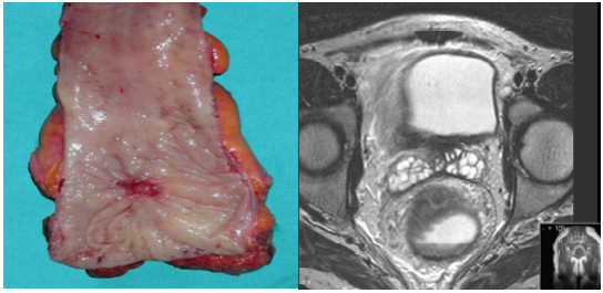
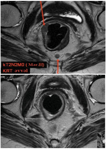
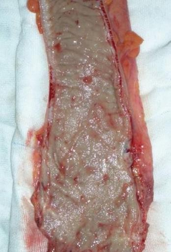
MRI scans of patients demonstrating a complete response to radiotherapy revealed total clinical regression of the tumor, with no visible residual mass observed (figure 2). Correspondingly, gross examination of the resected specimen confirmed the absence of macroscopic tumor involvement on the mucosal surface (figure 3), indicating a complete pathological response.
Although the efficacy of neoadjuvant radiotherapy in rectal cancer is well established particularly in enhancing resectability and reducing local recurrence its use is not without limitations. Preoperative chemoradiotherapy is a major contributor to postoperative complications, including anorectal dysfunction, alterations in sphincter function, and sexual dysfunction.
These disorders were manifested to varying degrees in 52 (65 %) of the patients in our study. We observed a significant decrease in both the resting and contractile pressures of the anal sphincter in these patients. Sexual dysfunction is a common complication following rectal cancer treatment. We believe that surgical trauma (damage to the erigent nerves), in addition to radiotherapy, plays a major role in the development of these complications.
Based on our observations and literature data, we recommend administering long-term radiochemotherapy (45-50 Gy, 225 mg/m² of 5-fluorouracil per day) prior to surgery in all eligible cases. The addition of chemotherapy to radiotherapy is intended to enhance the radiosensitivity of tumor cells. This combined approach helps minimize local recurrences. Following preoperative chemoradiotherapy, a waiting period of 6-8 weeks is recommended before proceeding with surgical intervention. It is crucial to perform a radical resection during surgery. We believe that the concept of correcting an inadequately performed total mesorectal excision with postoperative radiochemotherapy is, in our opinion, flawed. After surgical treatment, all patients underwent 4-6 courses of chemotherapy.
CONCLUSIONS
Preoperative radiotherapy is essential for rectal tumors staged as T1-T2 N-positive, T3 N0-1, and T4 N0-2.
For locally advanced rectal tumors (T3a N1-T4), the standard treatment involves surgical intervention combined with preoperative chemoradiotherapy.
Following long-term radiotherapy, in the laparoscopic group, 4 (18,2 %) patients exhibited complete radiosensitivity, 15 (68,2 %) patients showed partial radiosensitivity, and 3 (13,6 %) patients demonstrated radioresistance. In the open group, 5 (17,9 %) patients had complete radiosensitivity, 19 (67,9 %) had partial radiosensitivity, and 4 (14,3 %) displayed radioresistance (p=0,998).
Among patients with complete radiosensitivity (total of 9 patients), no local recurrence was observed in the first 3 years (0 %). In contrast, 5 (14,7 %) of partially radiosensitive patients and 7 (100 %) of radioresistant patients experienced local recurrence within the first 3 years.
Radiotherapy resulted in tumor regression as follows: in the laparoscopic group, 1st-degree regression was observed in 19,1±5,7 %, 2nd-degree regression in 51,1±7,3 %, 3rd-degree regression in 17,0±5,5 %, and 4th-degree regression in 12,8±4,9 %. In the open group, these values were 23,2±5,6 %, 44,6±6,6 %, 17,9±5,1 %, and 14,3±4,7 %, respectively.
In cases where the neoplasm is completely absorbed, a wait-and-see approach is not recommended. Total mesorectal excision should be performed after a period of 6-8 weeks.
References
- 1 Shaash, P., et al. Diverse strategies in locally advanced rectal cancer treatment. Journal of Clinical Oncology, 2025, Vol. 8.
- 2 Andrzej, P., et al. Rectal cancer update: Which treatment effects are the least “brutal”? International Journal of Radiation Oncology, Biology, Physics, 2024, Vol. 118, pp. 1-7.
- 3 Leila, T., et al. Analysis of radiation therapy quality assurance in NRG oncology RTOG 0848. International Journal of Radiation Oncology, Biology, Physics, 2024, Vol. 118, pp. 107-114.
- 4 Bbraendengen, M., Tvieit, K.M., Berglund, A., et al. Randomized phase III study comparing preoperative radiotherapy with chemoradiotherapy in non-resectable rectal cancers. Journal of Clinical Oncology, 2008, Vol. 26, pp. 3687.
- 5 Mohiuddin, M., Regine, W.F., John, W.J., et al. Preoperative chemoradiation in fixed distal rectal cancer: Dose-time factors for pathological complete response. International Journal of Radiation Oncology, Biology, Physics, 2000, Vol. 46, pp. 883.
- 6 Floris, S., et al. Compliance and toxicity of total neoadjuvant therapy for rectal cancer: A secondary analysis of the OPRA trial. International Journal of Radiation Oncology, Biology, Physics, 2024, Vol. 118, pp. 115-123.
- 7 Aaron, J., et al. Management of locally advanced rectal cancer: ASCO guideline. Journal of Clinical Oncology, 2024, Vol. 28.
- 8 Gerard, J.P., et al. Radiotherapy alone in the curative treatment of rectal carcinoma. The Lancet Oncology, 2003, Vol. 4, pp. 158-166.
- 9 Habr-Gama, A. Operative versus nonoperative treatment for stage 0 distal rectal cancer following chemoradiation therapy: Long-term results. Annals of Surgery, 2004, Vol. 4, pp. 711-717.
- 10 Medich, D., et al. Preoperative chemoradiotherapy and radical surgery for locally advanced distal rectal adenocarcinoma: Pathologic findings and clinical implications. Diseases of the Colon & Rectum, 2001, Vol. 44, pp. 1123-1128.
Declarations
Funding
None.
Conflict of interest
Authors declare that there is no conflict of interest. AUTHORSHIP CONTRIBUTION Conceptualization: E.H. Azimov, S Huseynov, A.A Ibrahimova. Data curation: E.H. Azimov, S Huseynov, A.A Ibrahimova. Formal analysis: E.H. Azimov, S Huseynov, A.A Ibrahimova. Drafting - original draft: E.H. Azimov, S Huseynov, A.A Ibrahimova. Writing - proofreading and editing: E.H. Azimov, S Huseynov, A.A Ibrahimova.
Authorship contributions
Drafting – original draft: E.H. Azimov, S Huseynov, A.A Ibrahimova.
Writing–review and editing: E.H. Azimov, S Huseynov, A.A Ibrahimova.