

Abstract
Bioactive ion-releasing restorative materials are marketed as an advance over inert resin composites because they release fluoride, calcium, phosphate or silicate ions intended to inhibit caries and promote remineralization. This narrative review examines the terminology, the laboratory and clinical evidence, and the controversies that separate those claims from measured performance. A documented search of PubMed, OpenAlex and Crossref identified studies published mainly between 2019 and 2026, supplemented by classic methodological references. The term bioactive is applied inconsistently; the FDI policy statement requires local, intended and nontoxic effects that do not compromise the restorative function. In vitro, glass ionomer cements release fluoride over time and can be recharged, whereas bioactive glasses, surface pre-reacted glass and calcium silicate fillers precipitate apatite in simulated body fluid, yet these surrogates do not predict clinical benefit. Clinical evidence supports a lower incidence of secondary caries for glass ionomer than for amalgam (risk ratio 0.20 in permanent teeth) and a similar incidence relative to resin composite, while glass ionomer and composite restorations show comparable survival in primary teeth and noncarious cervical lesions. Evidence for newer bioactive composites and alkasites remains short term and largely laboratory based. The central controversy is the translational gap: terminology and in vitro bioactivity outpace clinical data. Standardized terminology, surrogate validation and longer trials with caries-related outcomes are needed.
Keywords
dental materials, glass ionomer cements, dental caries, fluorides, remineralization, bioactive materials
INTRODUCTION
Secondary caries is the foremost reason for the failure and replacement of resin composite restorations, and its prevention remains the dominant challenge of adhesive restorative dentistry.(1,2) Conventional resin composites are essentially inert: they replace lost tissue but do not respond to the acidogenic biofilm that forms at the restoration margin. Because the mineral loss that characterizes recurrent lesions is driven by a persistent imbalance between demineralization and remineralization, materials that could release fluoride, calcium, phosphate or other ions into the adjacent tooth and biofilm have been proposed as a way of shifting that balance toward repair.(1,3) This rationale explains the rapid commercial expansion of products described as bioactive, smart or ion-releasing.
The terminology has, however, outpaced its definition. The term bioactive carries a positive connotation and is increasingly used in advertising, but clinicians, manufacturers and researchers do not share a single operational meaning, and the FDI World Dental Federation issued a policy statement to clarify the term and to warn against its overuse.(4,5) Parallel to that debate, a wide family of materials has accumulated laboratory and, to a lesser extent, clinical data: conventional and resin-modified glass ionomers, surface pre-reacted glass fillers, bioactive glass-loaded composites, calcium silicate cements and alkasites. The oldest member of the family, the glass ionomer cement, has released fluoride since its description in 1971.( 6) Their ion release profiles differ, and the laboratory surrogates used to demonstrate bioactivity, chiefly apatite formation in simulated body fluid, do not necessarily correspond to a measurable clinical benefit.( 7)
The objective of this review is to critically examine the evidence, the terminology and the controversies surrounding bioactive ion-releasing restorative materials. Three questions guide the synthesis: what definitions and classifications are in use; what does laboratory evidence show about ion release and its biological consequences; and what clinical evidence supports the caries-preventive and longevity claims made for these materials. The review adopts an evidence-matrix approach, quantifying how many and what kind of studies support each claim, so that the reader can weigh the strength of the evidence behind the marketing language.
METHODS
Design and reporting
This is a critical narrative review organized as an evidence matrix. It follows the SANRA items for narrative reviews and does not claim to be systematic or exhaustive: no PRISMA flow is reported, and selection was not intended to retrieve every eligible record.( 4) The scope was defined around direct restorative materials that release ions with a claimed biological effect on caries or remineralization; root canal sealers, pulp capping agents and orthodontic adhesives were considered only when they informed the mechanisms of ion release.
Sources and search
Searches were run through the project's bibliographic tool in PubMed, OpenAlex and Crossref. The core literature was restricted to the window 2019 to 2026 to reflect current evidence, and classic or methodological references published before that window were admitted when they defined the field, such as the original description of the glass ionomer cement(6) and the simulated body fluid method.(8) The query strings combined terms for the materials (glass ionomer, bioactive glass, calcium silicate, alkasite, giomer, surface pre-reacted glass), the phenomenon (ion release, fluoride release, apatite formation, remineralization) and the outcome (secondary caries, restoration survival). Retrieval was capped at forty records per query, and duplicates were removed by the tool.
Selection criteria
Records were retained when the title or abstract addressed ion release from a restorative material, its biological effect on enamel or dentin, or a clinical outcome of a restoration placed with such a material. Preference was given to original studies, randomized clinical trials and systematic reviews over narrative reviews when a factual claim about performance was cited. Studies were excluded when they concerned only endodontic sealers, bone cements or materials outside restorative dentistry, or when they were opinion pieces without data. No language restriction was applied, but all retained sources were published in English or Spanish.
Synthesis and quality appraisal
The extracted evidence was organized in a matrix with five thematic axes: terminology and classification, material families and ion release profiles, laboratory evidence, clinical evidence, and controversies. For each axis, claims were recorded together with the studies that support or contradict them, following a pre-registered internal template. Quality was graded by design, so that meta-analyses of randomized trials ranked above single trials, which ranked above laboratory studies; within laboratory evidence, standardized and long-term protocols were valued over single time-point measurements. The synthesis quantifies, where the sources allow it, how many sources underpin each claim, and it flags findings that rest on a single study or on a surrogate endpoint. Counts of supporting studies reported in the Development were taken from the retained records: a claim backed by a meta-analysis of 64 trials is described as such, and a claim resting on one laboratory study is identified as single-study evidence. Where a source reports its own included studies, that number is used rather than re-derived, so every count can be traced to the review or trial that produced it.
DEVELOPMENT
Evidence matrix
The retained evidence is summarized as a matrix that links each material family to the strongest study or review available for it, the design, the population or model, the principal finding and the level of evidence (Table 1). The matrix is the backbone of the synthesis that follows: each axis of the Development reads a row or a group of rows and weighs how many independent sources point in the same direction.
Study | Design | Population or model | Key finding | Evidence level |
|---|---|---|---|---|
Ge et al., 2023 | Meta-analysis of 64 trials | 8310 GIC and 5857 amalgam or composite restorations | Lower secondary caries for GIC than amalgam (RR 0.20 permanent); similar to composite | High |
Panetta et al., 2024 | Umbrella review of 13 systematic reviews | Primary and permanent teeth | GIC longevity varies by indication and material | High |
Bezerra et al., 2020 | Systematic review and meta-analysis | Noncarious cervical lesions | GIC and composite comparable in longevity | High |
Krishnakumar et al., 2024 | Systematic review of clinical trials | Primary teeth | HVGIC and composite comparable, with contradictory single studies | High |
Molina et al., 2019 | Randomized trial, 5 years | Persons with disability | ART/HVGIC and composite survival compared over 5 years | Moderate |
Garbim et al., 2024 | Randomized non-inferiority trial, 2 years | Primary molars, children aged 4 to 8 | Encapsulated GICs compared for ART survival and cost | Moderate |
Isikli et al., 2026 | Randomized trial, 18 months | Class II cavities, 50 patients | Alkasite and glass-hybrid comparable to composite on FDI criteria | Moderate |
Balkaya et al., 2019 | Randomized trial, 1 year | Class II cavities, 109 restorations | Bulk-fill, composite and reinforced GIC compared clinically | Moderate |
Marović et al., 2022 | In vitro with aging | Commercial ion-releasing materials | Giomer had the highest flexural values; alkasite outperformed GIC | Low |
Radwański et al., 2025 | In vitro | Commercial bioactive materials | Flexural strength ranged 86.6 to 120.4 MPa; one product had the lowest modulus and highest sorption | Low |
Aliberti et al., 2025 | In vitro, pH and temperature | Seven commercial materials | Ion release pH-, time- and temperature-dependent | Low |
Leão et al., 2021 | In vitro pH cycling | Enamel-material interface | S-PRG composite remineralized less than GIC but inhibited demineralization | Low |
Martínez-Llop et al., 2026 | Systematic review, 12 in vitro studies | Remineralizing resin composites | Ion release and apatite formation heterogeneous across formulations | Low |
Imazato et al., 2023 | Narrative review | S-PRG filler technology | Multiple-ion release associated with six in vitro bioactivities | Low |
Note: GIC = glass ionomer cement; HVGIC = high-viscosity glass ionomer cement; ART = atraumatic restorative treatment; RR = risk ratio; FS = flexural strength; S-PRG = surface pre-reacted glass ionomer; NCCL = noncarious cervical lesion. Evidence level reflects study design, not a formal GRADE rating. Source: Elaborated from the references cited in the text.
Terminology and classification of ion-releasing materials
The first controversy is semantic. The FDI policy statement records that bioactivity is used with a positive connotation in advertising, that the term means different things to clinicians, manufacturers and researchers, and that a restorative material should be called bioactive only when its effect is local, intended, nontoxic and does not interfere with the primary purpose of replacing dental tissue.(4) A companion analysis in the foundational dental literature reaches the same conclusion and adds that it is difficult to identify which measurable property actually qualifies a product as bioactive, which leaves the clinician unable to judge whether a bioactive product is clinically superior.(5) The definitions in circulation are collected in a synthesis table (Table 2).
Term | Working definition | Evidentiary basis |
|---|---|---|
Bioactive | Produces a local, intended, nontoxic effect without compromising the restoration | Consensus definition; no clinical validation required |
Biointeractive | Exchanges ions with the surrounding environment | Used interchangeably with bioactive; less frequent |
Smart material | Responds to an environmental stimulus | Commercial label; chemistries are heterogeneous |
Ion-releasing | Liberates measurable ions over time | Directly measurable in vitro |
Biomimetic | Reproduces tissue structure or mineralization | Experimental; no standardized test |
Note: SBF = simulated body fluid; ISO = International Organization for Standardization. Source: Elaborated from Schmalz et al., Ferracane, and the reviews cited in the text.
The same confusion extends to product families. A review of commercially available fluoride-releasing materials notes that formulations marketed as smart materials under several family names have few published studies and poorly explained chemistries, and it proposes a classification built on composition and setting reaction rather than on marketing labels.(9) A bibliometric review that screened 7161 records and retained 159 found the literature on bioactive materials for dentin coverage organized around overlapping descriptors such as ion-releasing, smart, biomimetic and smart replacement for dentin, which again points to an unsettled vocabulary.(10) One consequence is that the same product can be described as a giomer, an alkasite, a glass hybrid or a bioactive composite depending on the source, a situation that complicates any comparison of evidence across materials.(11)
The FDI statement grounds the debate by distinguishing the mechanisms through which a material may act and by cautioning that the positive connotation of the word is borrowed in advertising without the corresponding evidence.(4) The cost of that looseness appears in the research record, where the same chemistry is labelled with different terms and the bibliometric review recovered overlapping descriptors that fragment the field rather than organize it.(5,10) A related distinction, between bioactivity, remineralization and antibacterial action, is drawn in the comprehensive reviews but is not applied consistently across product literature, so a claim of bioactivity may refer to mineral precipitation in one paper and to bacterial inhibition in another.(11)
A constructive alternative to the marketing labels is a classification built on composition and setting reaction, as proposed for the fluoride-releasing families.( 9) Under such a scheme, a material is described first by its chemistry and only then by the property it claims, which would allow the evidence for each family to be pooled without the ambiguity introduced by proprietary names. The reviews that attempt this kind of taxonomy state that several of the newer formulations have few published studies and incompletely explained chemistries, so the taxonomy itself remains provisional.( 9)
Material families and ion release profiles
The oldest and best-characterized ion-releasing restorative family is the glass ionomer cement, formed from an ion-leachable glass and polyacrylic acid; its fluoride release is the reference against which newer products are measured.(12) Successive modifications, reviewed as chemically activated formulations with bioactive stimulation, have pursued remineralization, tissue integration and antimicrobial enhancement while retaining the basic acid-base chemistry of the parent material.(13) Glass ionomers release fluoride by an exchange mechanism in which the ion diffuses out of the set cement and can be reabsorbed from topical fluoride, so the material acts as a rechargeable reservoir rather than a one-off source.(14) This property, together with chemical adhesion and moisture tolerance, led the World Health Organization to add glass ionomer cement to its Model List of Essential Medicines in 2021, and clinical reviews describe its fluoride release as long lasting.(12) Resin-modified glass ionomers and the related compomer and giomer families release less fluoride than conventional glass ionomers, a trade-off that buys better aesthetics and handling at the cost of the ion reservoir.(9,15)
The second generation of ion-releasing products extends the chemistry beyond fluoride. Surface pre-reacted glass ionomer fillers liberate a set of ions, including fluoride, strontium, sodium, borate, aluminum and silicate, and the literature attributes six in vitro activities to this multi-ion release: tooth strengthening, acid neutralization, mineralization, microbial inhibition, matrix metalloproteinase inhibition and cell stimulation.(16) Bioactive glass-loaded resin composites release calcium, phosphate and silicate, raise the local pH and precipitate hydroxyapatite, with the effect increasing at higher filler loading.(17,18) Composites functionalized with hydrated calcium silicate follow the same logic, combining calcium release and apatite formation with antibacterial activity in experimental formulations.(19) The alkasite family adds an alkaline, acid-buffering behavior to fluoride and calcium release.(20,21)
The calcium silicate branch of the family illustrates how a chemistry migrates across indications. Apatite-forming calcium silicates originated in the pulp capping and endodontic lineage and have been adapted into restorative resin composites by adding hydrated calcium silicate filler, which imparts calcium release, antibacterial activity and apatite formation in experimental formulations.(19) The adaptation is promising because the apatite-forming chemistry is well established elsewhere, but the evidence for the restorative version remains confined to laboratory specimens, and the clinical history of the parent materials does not transfer automatically to a load-bearing composite.(7,19)
What the profiles share is a strong dependence on conditions and a predictable decay over time (Table 3). Across seven commercial materials, ion release varied with pH, temperature and exposure time, with the highest fluoride concentrations measured at acidic pH.( 22) Long-term assessment of contemporary materials showed that the giomer retained the highest flexural properties after aging while the alkasite outperformed a conventional glass ionomer, indicating that ion-releasing capability and mechanical durability are negotiated differently in each family.( 23)
Family | Main ions | Release pattern |
|---|---|---|
Conventional GIC | Fluoride, aluminum, sodium, calcium | Early burst, then sustained and rechargeable |
RMGIC | Fluoride, lower calcium | Lower and shorter than conventional GIC |
Giomer (S-PRG filler) | Fluoride, strontium, borate, silicate, aluminum | Multiple ions, sustained |
Bioactive glass composite | Calcium, phosphate, silicate, fluoride | pH-dependent, burst then decline |
Calcium silicate composite | Calcium, hydroxide | Alkaline, apatite-forming |
Alkasite | Fluoride, calcium, phosphate | Alkaline, acid-buffering |
Note: GIC = glass ionomer cement; RMGIC = resin-modified glass ionomer cement; S-PRG = surface pre-reacted glass ionomer. Patterns describe the dominant trend reported in the sources, not a single standardized measurement. Source: Elaborated from the reviews and in vitro studies cited in the text.
The mechanical cost of these formulations is uneven rather than universal. In one in vitro comparison, flexural strength ranged from 86.6 MPa to 120.4 MPa across commercial bioactive products, and the material with the lowest elastic modulus also showed the highest water sorption in ethanol and saliva, whereas a bulk-fill bioactive product reached the highest strength of the set.( 24) This dispersion means that no single mechanical profile can be attributed to the label bioactive, and the clinician must consult product-specific data instead of family-level claims.( 1)
The clinical workhorses of the family are the high-viscosity and encapsulated glass ionomers, whose handling properties were designed for atraumatic restorative treatment and for settings with limited isolation. Trials in primary molars have compared two encapsulated systems under atraumatic conditions and reported survival and cost outcomes over two years, and consensus guidance conditions the use of these materials on cavity size, load and caries risk.(25,26) That positioning matters for the review's central question, because the evidence base of the bioactive label rests on these fluoride-releasing cements rather than on the multi-ion composites that carry the newer marketing claims.
Laboratory evidence
Laboratory bioactivity is almost always demonstrated with the simulated body fluid method, in which apatite precipitation on a surface is read as evidence of bioactivity, a surrogate originally developed for bone-bonding materials.(7) The method is standardized but its predictive value for the mouth is weak, because the intraoral environment differs from acellular simulated body fluid in pH, protein content and fluid flow, and because apatite precipitation does not by itself demonstrate that enamel or dentin is protected or repaired.(7,17) Resin composites functionalized with bioactive glass illustrate the point: they release calcium and phosphate and precipitate hydroxyapatite in vitro, yet the clinical meaning of that precipitation has not been established.(17,18)
Within the laboratory domain, the evidence is internally consistent on three points. Fluoride-releasing materials resist enamel demineralization and support remineralization under pH cycling; a comparative study of a glass ionomer, a resin-modified glass ionomer and an alkasite found that the alkasite raised pH initially and that all three released fluoride and calcium, with the glass ionomer showing the strongest demineralization resistance.(21) A prereacted glass filler composite inhibited enamel demineralization but remineralized less than a glass ionomer, a result that separates the two properties and cautions against equating any fluoride release with full remineralizing capacity.(27) Bioactive glasses promote apatite formation and raise pH, but conventional 45S5 glass is mechanically weak and prone to an initial burst of ion release, which has motivated secondary oxide, hybrid and mesoporous formulations whose oral durability remains unproven.(7)
The experimental composite literature is larger than its clinical counterpart but heterogeneous. A systematic review restricted to in vitro studies of remineralizing resin composites retained 12 studies and found that ion release efficacy, bioactive filler composition and mechanical properties varied so widely that no formulation could be singled out as superior.(28) Experimental composites with hydrated calcium silicate showed dose-dependent antibacterial and apatite-forming behavior in one study.(19) For the wider remineralization field, systematic reviews of enamel remineralization techniques and of casein phosphopeptide-amorphous calcium phosphate, nanohydroxyapatite and bioactive glass against fluoride converge on a shared conclusion: fluoride remains the benchmark, and the nonfluoride agents show promise but inconsistent depth and durability of remineralization.(29-31) This body of work also confirms a basic biochemical constraint: net remineralization requires an adequate supply of calcium and phosphate, and saliva is the principal reservoir that delivers these ions to the enamel surface.(3)
Beyond mineral effects, the multi-ion products are evaluated for antibacterial activity. The surface pre-reacted glass filler is credited in the literature with several in vitro activities, among them microbial and fungal inhibition and matrix metalloproteinase inhibition, and experimental calcium silicate composites showed antibacterial behavior alongside apatite formation.(16,19) Alkaline and acid-buffering effects are measured with the same logic: several bioactive formulations raise the local pH, which is proposed to offset the acid attack of the biofilm, although the effect is again demonstrated in solution rather than in a clinical lesion.(20,21) The result is a laboratory profile with three overlapping claims, mineral deposition, pH buffering and microbial inhibition, none of which has yet been tied to a reduction in recurrent caries in a controlled trial.
The heterogeneity of these protocols limits what the laboratory evidence can say as a whole. Studies differ in immersion medium, specimen geometry, aging, measurement time points and the units used to report release, so the field produces many single comparisons and few cumulative statements.(22,23) The systematic review of in vitro remineralizing composites encountered exactly this problem, finding that ion release efficacy and mechanical properties varied across 12 studies in ways that prevented a ranking of formulations.(28) Standardization of the release assay, from specimen conditioning to the reporting of concentration over time, is therefore a precondition for any future claim that one bioactive product outperforms another.
Clinical evidence
Clinical data are concentrated in the glass ionomer family and are strongest for the outcome that matters most, secondary caries (Table 4). A meta-analysis of 64 randomized trials pooling 8310 glass ionomer restorations against 5857 amalgam or composite restorations found a lower incidence of secondary caries for glass ionomer than for amalgam in primary teeth (risk ratio 0.55; 95% CI 0.41 to 0.72) and in permanent teeth (risk ratio 0.20; 95% CI 0.11 to 0.38), while the incidence relative to resin composite was similar in both dentitions (risk ratio 0.92 and 0.77, respectively).(32) The same review found no difference between conventional and resin-modified glass ionomers for secondary caries. This is the single most informative result of the field: the fluoride-releasing material is cariostatic compared with amalgam and roughly equivalent to composite, which frames bioactivity as a protective property rather than a guarantee of superiority over modern composites.
The precision of that estimate deserves attention. The meta-analysis pooled trials with follow-up ranging from one to ten years, and the confidence interval for the permanent-dentition comparison against composite was wide (risk ratio 0.77; 95% CI 0.39 to 1.51), so the equivalence between glass ionomer and composite for secondary caries in permanent teeth is imprecise rather than firmly established.(32) Against amalgam the advantage was precise and consistent across dentitions, which is the comparison in which the fluoride-releasing mechanism should show its effect if it is real. The contrast between the two comparisons, one precise and one wide, is a reminder that the cariostatic benefit is demonstrated against a non-fluoridated control and is not yet established as an advantage over modern resin composites.
Survival data tell a consistent story. An umbrella review of 13 systematic reviews of glass ionomer longevity in primary and permanent teeth concluded that survival varies by indication and by the specific glass ionomer formulation, without a uniform advantage over composite.(33) A systematic review of clinical trials in primary teeth likewise found high-viscosity glass ionomer and composite comparable, with isolated contradictory studies.(34) In noncarious cervical lesions, a meta-analysis found glass ionomer and composite comparable in clinical performance and longevity.(35) For atraumatic restorative treatment, the evidence favors acceptable rather than superior performance: a 5-year randomized comparison in persons with disability compared high-viscosity glass ionomer ART restorations with composite, and a 2-year non-inferiority trial in primary molars compared two encapsulated glass ionomers under ART conditions.(25,36) A systematic review of ART in elderly patients, based on seven eligible studies, reported survival that supports the approach in that population without resolving which glass ionomer performs best.(37)
The clinical literature is also distinctive for where it was obtained. The strongest survival data come from populations with limited access or with special needs: a five-year comparison in persons with disability, a two-year non-inferiority trial in schoolchildren, and a systematic review of ART in elderly patients.(25,36,37) This is a strength in external validity, because these are the patients for whom a moisture-tolerant, fluoride-releasing material is most useful, but it also means the evidence reflects atraumatic cavity preparation and selective caries removal more than conventional Class II composite placements. The clinician extrapolating these results to load-bearing posterior restorations in healthy adults is therefore reading beyond the populations that generated the data.
The newer families have only short-term clinical data. An 18-month randomized trial in 50 patients compared an alkasite and a glass hybrid with a resin composite in Class II cavities and scored the three materials with FDI criteria, providing the first controlled comparison for this generation but no evidence beyond a year and a half.(38) A one-year randomized study compared a reinforced glass ionomer with a bulk-fill composite and a conventional composite in Class II cavities,(39) and a two-year split-mouth study compared a coated glass ionomer with a bulk-fill composite in Class I and II cavities.(40) These trials are consistent with the older literature on composite longevity, which reports high survival for posterior composites in the range of several years, so a new bioactive material must at least match that benchmark to be justified.(41,42) Consensus guidance on glass ionomer thresholds stresses that the decision to use these materials depends on cavity size, load and caries risk, which again places the clinical value of ion release in a specific context rather than in the material label.(26)
Outcome | Comparison | Direction of evidence | Level |
|---|---|---|---|
Secondary caries | GIC vs amalgam | Lower for GIC (RR 0.55 primary; 0.20 permanent) | High, meta-analysis of 64 RCTs |
Secondary caries | GIC vs composite | Similar (RR 0.92 primary; 0.77 permanent) | High, meta-analysis of 64 RCTs |
Survival, primary teeth | HVGIC vs composite | Comparable, with contradictory single studies | High to moderate |
Survival, NCCL | GIC vs composite | Comparable | High, meta-analysis |
Survival, ART | Encapsulated HVGIC systems | Acceptable at 2 to 5 years | Moderate |
Short-term performance | Alkasite and glass-hybrid vs composite | Comparable at 18 months | Moderate, single RCT |
Note: GIC = glass ionomer cement; HVGIC = high-viscosity glass ionomer cement; ART = atraumatic restorative treatment; RR = risk ratio; CI = confidence interval; NCCL = noncarious cervical lesion; RCT = randomized clinical trial. Levels reflect the design of the underlying evidence. Source: Elaborated from the meta-analyses and trials cited in the text.
Controversies and evidence gaps
The central controversy is translational. Laboratory surrogates, apatite formation in particular, are abundant, but no trial has yet shown that a composite or alkasite marketed as bioactive reduces secondary caries or extends survival beyond what a conventional composite achieves.(4,43) The resin literature is explicit that secondary caries remains the leading cause of composite failure and that the main challenge of bioactive resin development is to translate ion release into protection of the bonded interface, a goal that remains unmet.(1) Bioactive glass reviews state the same limitation in the opposite direction: the materials are reactive and antibacterial in vitro, but their clinical translation is limited and their durability under oral conditions is not established.(7) The gap can be represented as a chain from the measured property to the patient outcome (Figure 1).
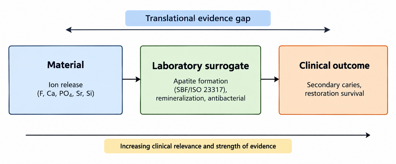
Figure 1. Chain from ion release to clinical outcome and the translational evidence gap.
Note: SBF = simulated body fluid; ISO = International Organization for Standardization.A second, related gap is the asymmetry between terminology and measurement. Products are named for a mechanism, but the clinician can only observe outcomes, and the published record connects the two mainly within the glass ionomer family.( 5) The classification problem documented for fluoride-releasing families has not been resolved by the newer multi-ion products, which arrive under names that describe neither their chemistry nor their evidence.(9,10) This asymmetry is aggravated by comparison with nonrestorative ion-releasing agents. Silver diamine fluoride arrests caries within hours of application and is supported by a growing clinical literature, and topical bioactive glasses and calcium phosphate systems have been evaluated against fluoride for initial lesions; the restorative materials discussed here do not yet carry evidence of that maturity.( 30,31,44)
A further source of confusion is the difference between restorative and non-restorative ion delivery. A restorative material must release ions from a bulk that is also expected to bear load for years, whereas a topical agent can be reapplied, which is why silver diamine fluoride and toothpaste-based fluoride have reached a maturity of clinical evidence that the restorative bioactives have not.(44,45) Recognizing this difference would discipline the vocabulary, because a material that releases fluoride for a few weeks is not equivalent to a topical regimen that replenishes the ion at every application.
The third gap is the durability of the ion reservoir and of the material itself. Fluoride release from glass ionomers is rechargeable but declines between exposures,(14) and the burst-and-decay pattern of bioactive glass formulations raises the question of whether protection lasts as long as the restoration is expected to.(7) Mechanical data add a further caveat: some bioactive products absorb more water and flex less than a conventional composite, so the clinician may trade structural performance for an unproven biological benefit.(24) These three gaps, the missing trial, the terminology gap and the durability question, define the current state of the field.
DISCUSSION
The principal finding of this matrix-based synthesis is that the strength of the clinical evidence is concentrated in the oldest family and thinnest in the newest one. Glass ionomer cements carry a meta-analysis of 64 randomized trials showing less secondary caries than amalgam and similar caries to composite, whereas the products most aggressively marketed as bioactive, the multi-ion composites and alkasites, rest on short-term trials and laboratory surrogates.(32,38) The evidence is thus inversely related to the novelty of the label, and the term bioactive currently signals a mechanism more than a demonstrated clinical advantage.
Graded by design, the findings separate into three tiers. The first tier, secondary caries prevention by glass ionomer, rests on a meta-analysis of randomized trials and is the only finding that would survive a formal certainty assessment unchanged. The second tier, restoration survival, rests on meta-analyses and trials of variable follow-up, and its conclusions are comparable rather than superior. The third tier, the multi-ion activity of the newer products, rests on laboratory studies and short trials, and any claim derived from it should be read as hypothesis rather than established fact.( 32,33,38) This tiering, not the word bioactive, is the information a clinician needs when choosing a material.
Across the clinical outcomes the direction of evidence is consistent. Secondary caries, survival in primary teeth, noncarious cervical lesion longevity and ART performance all converge on the same picture: glass ionomers protect against caries at least as well as composite and are comparable rather than superior in longevity.(32-35,37) The level of evidence is highest for the caries outcome, because it is the only one pooled across many trials with a narrow confidence interval, and moderate for survival, where single studies contradict each other.(34) This consistency is reassuring but it also sets a high bar: a new bioactive material must beat a composite on a caries-related endpoint, and the headroom is small because composite already performs close to glass ionomer for secondary caries in the meta-analysis.(32)
The laboratory and clinical literatures diverge in a way that deserves explanation rather than dismissal. In vitro, the differences between materials are large and reproducible: glass ionomers resist demineralization more than resin-modified variants, a prereacted glass composite remineralizes less than a glass ionomer, and bioactive glasses precipitate apatite that composite does not.( 17,21,27) Clinically, these same materials look alike. The most plausible explanation is that the surrogate endpoints measure a single mechanism under idealized conditions, whereas secondary caries and survival depend also on the operator, the cavity, the patient's caries risk and the integrity of the adhesive interface, factors that dilute a laboratory advantage.(1,41) The simulated body fluid assay, designed for bone-bonding implants, is a weak model of a biofilm-driven lesion at a restoration margin, which is why apatite formation has not translated into fewer recurrent lesions.(8)
The mechanisms that are well established support a cautious interpretation. Fluoride release from glass ionomers is rechargeable and acts locally by inhibiting demineralization and accelerating remineralization, and the multi-ion release of surface pre-reacted glass adds pH buffering and matrix metalloproteinase inhibition in vitro.(14,16) These are plausible protective pathways, and they align with the fluoride literature, which identifies a sustained low fluoride concentration at the biofilm as the decisive condition for caries prevention.(45) Calcium and phosphate release faces a harder biochemical test, because net remineralization is limited by ion availability in saliva and depends on reconstructing the collagen-apatite hierarchy of dentin rather than on precipitating mineral alone.(3,28,46) The distinction explains why fluoride-releasing materials have clinical data and calcium-releasing composites do not.
The applicability of these findings differs by dentition. The secondary caries advantage of glass ionomer over amalgam was present in both dentitions, but the survival comparisons in primary teeth showed contradictory results across studies, and the umbrella review concluded that longevity depends on the specific formulation and indication.(32-34) In permanent posterior teeth, where composite is the first choice, the clinical question is not whether a bioactive material is better than amalgam but whether it is better than composite, and that question remains open.(42) The operative consequence is that the choice between a glass ionomer and a composite should be driven by caries risk, isolation and cavity size rather than by a bioactivity label.(26)
Compared with previous reviews, this synthesis reaches a more conservative conclusion. Recent comprehensive reviews describe the mechanisms, assessment methods and clinical applications of bioactive materials and frame them as a paradigm shift toward functional restorations.(11,43) The present review shares that reading of the mechanisms but weights the evidence differently: a paradigm shift requires outcome data, and outcome data exist only for the fluoride-releasing families. Reviews of bioactive glass and of remineralizing agents reach the same boundary from the materials side, acknowledging that clinical translation remains limited.(7,31) The bibliometric record shows why the discrepancy persists, because most of the published work is laboratory based and the vocabulary is not standardized, which makes evidence difficult to aggregate.( 10)
Several limitations temper these conclusions. The review is narrative rather than systematic, so relevant studies may have been missed and the selection depended on the judgment of one reader, a limitation that a prospective protocol with dual screening and a registered search would address. The synthesis relied partly on abstracts when full texts were not openly available, which can overstate or understate a study's results; a full-text extraction with risk-of-bias assessment would correct this. The clinical evidence for the newer materials is short term, so the review cannot exclude a benefit that appears only after several years; longer trials with caries as the primary outcome would resolve it. The mechanical and ion release data come from heterogeneous in vitro protocols, which limits direct comparison across brands; standardized reporting of ion concentration, time course and specimen conditioning would make the laboratory evidence cumulative. Finally, the quality labels in the matrix reflect study design rather than a formal GRADE rating, and a formal certainty-of-evidence assessment per outcome would strengthen the conclusions.
For clinical practice, the implications follow the evidence. In patients at high caries risk, or where isolation is compromised, a glass ionomer or high-viscosity glass ionomer ART restoration is a defensible choice because its cariostatic property is documented at the level of a meta-analysis of trials.(32,36) For the multi-ion composites and alkasites, the clinician should treat them as composites with a plausible but unproven added benefit and should not expect the label bioactive to substitute for caries risk control, which remains the stronger determinant of restoration survival.(26,42) The honest position, consistent with the FDI policy statement, is to require from any bioactive product the same outcome evidence demanded of a new composite.(4)
The research agenda follows from the gaps identified. Trials that randomize a bioactive composite or alkasite against a conventional composite in caries-active patients, with recurrent caries as the primary outcome and follow-up beyond three years, would directly test the translational claim that laboratory apatite formation predicts fewer recurrent lesions. Standardized reporting of ion concentration, time course and specimen conditioning would make the laboratory evidence cumulative rather than descriptive, and a validated surrogate that links ion release to enamel protection under biofilm conditions would bridge the two literatures. Consensus terminology, applied at the level of journals and regulatory dossiers, would do the same for the vocabulary, so that a future reader can tell from the title whether a study measured an ion, a surrogate or a clinical outcome.( 4,7)
CONCLUSIONS
The review set out to weigh the evidence, the terminology and the controversies of bioactive ion-releasing restorative materials, and the evidence supports four conclusions, one for each objective. The terminology was found to be inconsistent, and the FDI definition of bioactivity as a local, intended and nontoxic effect that does not compromise the restoration offers the most defensible standard. The laboratory evidence showed that glass ionomers release fluoride in a sustained and rechargeable manner while bioactive glasses, surface pre-reacted glass and calcium silicate fillers precipitate apatite and raise pH, although these surrogates do not predict clinical benefit. The clinical evidence demonstrated a lower incidence of secondary caries for glass ionomer than for amalgam, a similar incidence relative to composite, and comparable survival across indications, whereas the newer bioactive composites and alkasites carried only short-term data. The review identified a persistent gap between marketing terminology and measured performance, with the missing clinical trial as the field's central unresolved question. Until ion-releasing materials are tested against composite on caries-related outcomes, the term bioactive should be read as a description of mechanism, not as evidence of superiority.
References
- 1 Melo MAS, Garcia IM, Mokeem L, Weir MD, Xu HHK, Montoya C, et al. Developing Bioactive Dental Resins for Restorative Dentistry. J Dent Res. 2023;102(11):1180-1190. doi: 10.1177/00220345231182357
- 2 German MJ. Developments in resin-based composites. Br Dent J. 2022;232(9):638-643. doi: 10.1038/s41415-022-4240-8
- 3 Farooq I, Bugshan A. The role of salivary contents and modern technologies in the remineralization of dental enamel: a narrative review. F1000Res. 2021;9:171. doi: 10.12688/f1000research.22499.3
- 4 Schmalz G, Hickel R, Price RB, Platt JA. Bioactivity of Dental Restorative Materials: FDI Policy Statement. International Dental Journal. 2023;73(1):21-27. doi: 10.1016/j.identj.2022.11.012
- 5 Ferracane JL, Sidhu SK, Melo MAS, Yeo ISL, Diogenes A, Darvell BW. Bioactive dental materials. JADA Foundational Science. 2023;2:100022. doi: 10.1016/j.jfscie.2023.100022
- 6 Wilson AD, Kent BE. The glass‐ionomer cement, a new translucent dental filling material. J Appl Chem. 1971;21(11):313-313. doi: 10.1002/jctb.5020211101
- 7 Farjaminejad R, Farjaminejad S, Garcia-Godoy F, Jalali M. The Role of Bioactive Glasses in Caries Prevention and Enamel Remineralization. Applied Sciences. 2025;15(24):13157. doi: 10.3390/app152413157
- 8 Kokubo T, Yamaguchi S. Novel bioactive materials developed by simulated body fluid evaluation: Surface-modified Ti metal and its alloys. Acta Biomaterialia. 2016;44:16-30. doi: 10.1016/j.actbio.2016.08.013
- 9 Francois P, Fouquet V, Attal JP, Dursun E. Commercially Available Fluoride-Releasing Restorative Materials: A Review and a Proposal for Classification. Materials. 2020;13(10):2313. doi: 10.3390/ma13102313
- 10 Pires PM, Rosa TDC, Ribeiro-Lages MB, Duarte ML, Cople Maia L, Neves ADA, et al. Bioactive Restorative Materials Applied over Coronal Dentine—A Bibliometric and Critical Review. Bioengineering. 2023;10(6):731. doi: 10.3390/bioengineering10060731
- 11 Abozaid D, Azab A, Bahnsawy MA, Eldebawy M, Ayad A, soomro R, et al. Bioactive restorative materials in dentistry: a comprehensive review of mechanisms, clinical applications, and future directions. Odontology. 2026;114(2):349-377. doi: 10.1007/s10266-025-01162-w
- 12 Ge KX, Yu-Hang Lam W, Chu CH, Yu OY. Updates on the clinical application of glass ionomer cement in restorative and preventive dentistry. Journal of Dental Sciences. 2024;19(Suppl 1):S1-S9. doi: 10.1016/j.jds.2024.07.021
- 13 Makanjuola J, Deb S. Chemically Activated Glass-Ionomer Cements as Bioactive Materials in Dentistry: A Review. Prosthesis. 2023;5(1):327-345. doi: 10.3390/prosthesis5010024
- 14 Nicholson JW, Sidhu SK, Czarnecka B. Fluoride exchange by glass-ionomer dental cements and its clinical effects: a review. Biomaterial Investigations in Dentistry. 2023;10(1):2244982. doi: 10.1080/26415275.2023.2244982
- 15 Porenczuk A, Jankiewicz B, Naurecka M, Bartosewicz B, Sierakowski B, Gozdowski D, et al. A comparison of the remineralizing potential of dental restorative materials by analyzing their fluoride release profiles. Adv Clin Exp Med. 2019;28(6):815-823. doi: 10.17219/acem/94140
- 16 Imazato S, Nakatsuka T, Kitagawa H, Sasaki JI, Yamaguchi S, Ito S, et al. Multiple-Ion Releasing Bioactive Surface Pre-Reacted Glass-Ionomer (S-PRG) Filler: Innovative Technology for Dental Treatment and Care. JFB. 2023;14(4):236. doi: 10.3390/jfb14040236
- 17 Yun J, Burrow MF, Matinlinna JP, Wang Y, Tsoi JKH. A Narrative Review of Bioactive Glass-Loaded Dental Resin Composites. JFB. 2022;13(4):208. doi: 10.3390/jfb13040208
- 18 Par M, Gubler A, Attin T, Tarle Z, Tarle A, Tauböck TT. Ion release and hydroxyapatite precipitation of resin composites functionalized with two types of bioactive glass. Journal of Dentistry. 2022;118:103950. doi: 10.1016/j.jdent.2022.103950
- 19 Yang SY, Han AR, Choi JW, Kim KM, Kwon JS. Novel antibacterial and apatite forming restorative composite resin incorporated with hydrated calcium silicate. Biomater Res. 2023;27(1):25. doi: 10.1186/s40824-023-00364-z
- 20 Kasraei S, Haghi S, Valizadeh S, Panahandeh N, Nejadkarimi S. Phosphate Ion Release and Alkalizing Potential of Three Bioactive Dental Materials in Comparison with Composite Resin. International Journal of Dentistry. 2021;2021:1-8. doi: 10.1155/2021/5572569
- 21 Kim MJ, Lee MJ, Kim KM, Yang SY, Seo JY, Choi SH, et al. Enamel Demineralization Resistance and Remineralization by Various Fluoride-Releasing Dental Restorative Materials. Materials. 2021;14(16):4554. doi: 10.3390/ma14164554
- 22 Aliberti A, Di Duca F, Triassi M, Montuori P, Scippa S, Piscopo M, et al. The Effect of Different pH and Temperature Values on Ca2+, F−, PO43−, OH−, Si, and Sr2+ Release from Different Bioactive Restorative Dental Materials: An In Vitro Study. Polymers. 2025;17(5):640. doi: 10.3390/polym17050640
- 23 Marovic D, Par M, Posavec K, Marić I, Štajdohar D, Muradbegović A, et al. Long-Term Assessment of Contemporary Ion-Releasing Restorative Dental Materials. Materials. 2022;15(12):4042. doi: 10.3390/ma15124042
- 24 Radwanski M, Zmyslowska-Polakowska E, Osica K, Krasowski M, Sauro S, Hardan L, et al. Mechanical properties of modern restorative “bioactive” dental materials - an in vitro study. Sci Rep. 2025;15(1):3552. doi: 10.1038/s41598-025-86595-7
- 25 Garbim JR, Saihara CS, Olegário IC, Hesse D, Araujo MP, Bonifácio CC, et al. 2-year survival and cost analysis of occlusoproximal ART restorations using encapsulated glass ionomer cement in primary molars: a randomized controlled trial. BMC Oral Health. 2024;24(1):647. doi: 10.1186/s12903-024-04357-9
- 26 de Lima Navarro MF, Pascotto RC, Borges AFS, Soares CJ, Raggio DP, Rios D, et al. Consensus on glass-ionomer cement thresholds for restorative indications. Journal of Dentistry. 2021;107:103609. doi: 10.1016/j.jdent.2021.103609
- 27 Leão I, Araújo N, Scotti C, Mondelli R, de Amoêdo Campos Velo M, Bombonatti J. The Potential of a Bioactive, Pre-reacted, Glass-Ionomer Filler Resin Composite to Inhibit the Demineralization of Enamel in Vitro. Operative Dentistry. 2021;46(1):E11-E20. doi: 10.2341/19-151-l
- 28 Martínez-Llop A, Sanz JL, Folguera S, Villanueva-Castellote Á, Paredes A, Ghilotti J. Remineralizing Composites: A Systematic Review of In Vitro Studies. J Funct Biomater. 2026;17(11):536. doi: 10.3390/jfb17110536
- 29 Malcangi G, Patano A, Morolla R, De Santis M, Piras F, Settanni V, et al. Analysis of Dental Enamel Remineralization: A Systematic Review of Technique Comparisons. Bioengineering. 2023;10(4):472. doi: 10.3390/bioengineering10040472
- 30 Santhosh VN, Ankola AV, Sankeshwari RM, Varghese AS, Ragu K, Parimala YK, et al. Remineralization Potential of Casein Phosphopeptide–Amorphous Calcium Phosphate, Nanohydroxyapatite Crystals, and Bioactive Glass on Initial Enamel Lesions: A Systematic Review and Meta-analysis. International Journal of Clinical Pediatric Dentistry. 2025;18(5):560-572. doi: 10.5005/jp-journals-10005-3129
- 31 Dai LL, Mei ML, Chu CH, Lo ECM. Mechanisms of Bioactive Glass on Caries Management: A Review. Materials. 2019;12(24):4183. doi: 10.3390/ma12244183
- 32 Ge KX, Quock R, Chu CH, Yu OY. The preventive effect of glass ionomer cement restorations on secondary caries formation: A systematic review and meta-analysis. Dental Materials. 2023;39(12):e1-e17. doi: 10.1016/j.dental.2023.10.008
- 33 Panetta A, Lopes P, Novaes TF, Rio R, Fernandes GVO, Mello-Moura ACV. Evaluating Glass Ionomer Cement Longevity in the Primary and Permanent Teeth—An Umbrella Review. JFB. 2024;15(2):48. doi: 10.3390/jfb15020048
- 34 Krishnakumar K, Kalaskar R, Kalaskar A, Bhadule S, Joshi S. Clinical Effectiveness of High-viscosity Glass Ionomer Cement and Composite Resin as a Restorative Material in Primary Teeth: A Systematic Review of Clinical Trials. International Journal of Clinical Pediatric Dentistry. 2024;17(2):221-228. doi: 10.5005/jp-journals-10005-2757
- 35 Bezerra IM, Brito ACM, de Sousa SA, Santiago BM, Cavalcanti YW, de Almeida LDFD. Glass ionomer cements compared with composite resin in restoration of noncarious cervical lesions: A systematic review and meta-analysis. Heliyon. 2020;6(5):e03969. doi: 10.1016/j.heliyon.2020.e03969
- 36 MOLINA GF, FAULKS D, MULDER J, FRENCKEN JE. High-viscosity glass-ionomer vs. composite resin restorations in persons with disability: Five-year follow-up of clinical trial. Braz oral res. 2019;33:e099. doi: 10.1590/1807-3107bor-2019.vol33.0099
- 37 Maia TS, Lima TD, Vieira WDA, Blumenberg C, Agostini BA, Nascimento GG, et al. Survival of atraumatic restorative treatment restorations in the elderly patients: a systematic review. Braz oral res. 2021;35:e130. doi: 10.1590/1807-3107bor-2021.vol35.0130
- 38 Işıklı K, Koc Vural U, Yalcin Cakir F. Clinical performance of alkasite and glass-hybrid restorations compared with resin composite in class II cavities: a randomized clinical trial. Clin Oral Invest. 2026;30(5):211. doi: 10.1007/s00784-026-06869-w
- 39 Balkaya H, Arslan S, Pala K. A randomized, prospective clinical study evaluating effectiveness of a bulk-fill composite resin, a conventional composite resin and a reinforced glass ionomer in Class II cavities: one-year results. J Appl Oral Sci. 2019;27:e20180678. doi: 10.1590/1678-7757-2018-0678
- 40 Uzel İ, Aykut-Yetkiner A, Ersin N, Ertuğrul F, Atila E, Özcan M. Evaluation of Glass-Ionomer versus Bulk-Fill Resin Composite: A Two-Year Randomized Clinical Study. Materials. 2022;15(20):7271. doi: 10.3390/ma15207271
- 41 Josic U, D’Alessandro C, Miletic V, Maravic T, Mazzitelli C, Jacimovic J, et al. Clinical longevity of direct and indirect posterior resin composite restorations: An updated systematic review and meta-analysis. Dental Materials. 2023;39(12):1085-1094. doi: 10.1016/j.dental.2023.10.009
- 42 Pizzolotto L, Moraes RR. Resin Composites in Posterior Teeth: Clinical Performance and Direct Restorative Techniques. Dentistry Journal. 2022;10(12):222. doi: 10.3390/dj10120222
- 43 Sauro S, Carvalho RM, Ferracane J. The rise of advanced bioactive restorative materials: Are they redefining operative dentistry?. Dental Materials. 2025;41(11):1411-1429. doi: 10.1016/j.dental.2025.08.003
- 44 Jamal D, AlMushayt A, Abujamel T, Bamashmous S, Bamashmous N, Alamoudi N. Silver diamine fluoride: the science behind the action – a narrative review. BMC Oral Health. 2025;25(1):1195. doi: 10.1186/s12903-025-06107-x
- 45 Šutej I, Bašić K, Peroš K. The Cariostatic Mechanisms of Fluoride—An Updated Review. Dentistry Journal. 2026;14(7):390. doi: 10.3390/dj14070390
- 46 Reynolds E. Calcium phosphate‐based remineralization systems: scientific evidence?. Australian Dental Journal. 2008;53(3):268-273. doi: 10.1111/j.1834-7819.2008.00061.x
Declarations
Funding
No financing
Conflict of interest
None
Authorship contributions
Drafting – original draft: Arlin Castro Diaz.
Writing–review and editing: Arlin Castro Diaz.