

Abstract
Introduction: Scaling and root planing is the cornerstone of non-surgical therapy for stage III-IV periodontitis, but its results in deep pockets are incomplete. Antimicrobial photodynamic therapy has been proposed as an adjunct, although its added benefit remains uncertain. Methods: Randomized clinical trials comparing scaling and root planing alone with scaling and root planing plus antimicrobial photodynamic therapy in adults with stage III-IV or severe periodontitis were searched in OpenAlex, PubMed and Crossref. Probing depth reduction and clinical attachment gain were pooled with a random-effects model, and risk of bias was assessed with the Cochrane RoB 2 tool. Results: Ten trials were included. At three months, adjunctive photodynamic therapy produced a greater probing depth reduction than scaling and root planing alone (mean difference 0.77 mm; 95% CI 0.46 to 1.09; I2 = 64%), whereas the gain in clinical attachment was not statistically significant (mean difference 0.72 mm; 95% CI -0.29 to 1.74; I2 = 89%). Conclusions: Adjunctive antimicrobial photodynamic therapy provides a modest additional reduction in probing depth at three months, without a demonstrable gain in clinical attachment, in stage III-IV periodontitis. The evidence is limited by small trials and marked heterogeneity.
Keywords
photochemotherapy, periodontitis, dental scaling, root planing, systematic review, meta-analysis
INTRODUCTION
Periodontitis is a chronic multifactorial inflammatory disease initiated by a dysbiotic subgingival biofilm and sustained by an exaggerated host response, which progressively destroys the periodontal ligament, the root cementum and the alveolar bone and constitutes one of the leading causes of tooth loss in adults.(1) Severe periodontitis affects approximately 11% of the adult population worldwide, which makes it one of the most prevalent chronic diseases of humanity, and its functional and nutritional consequences extend well beyond the oral cavity, because tooth loss compromises mastication, speech and quality of life. The 2017 World Workshop replaced the former diagnostic labels of chronic and aggressive periodontitis with a staging and grading system that links severity and complexity to treatment. In this framework, stages III and IV identify the severe forms: probing depths of 6 mm or more, interdental attachment loss of at least 5 mm, radiographic bone loss extending to the middle or apical third of the root, and, in stage IV, the loss of five or more teeth attributable to periodontitis together with the need for complex rehabilitation.(2,3) It is precisely in these stages that non-surgical therapy becomes least predictable. Subgingival instrumentation can remove biofilm and calculus from the accessible root surface, but its mechanical access is limited at the base of deep pockets, in furcation entrances and in root concavities, so residual deposits and bacterial recolonization are common and the treatment outcome is determined in part by the initial probing depth.(4,5) Deep residual pockets that persist after instrumentation are the sites at which the disease is most likely to progress, and they are also the sites at which an adjunctive local decontamination would be expected to add the most. The persistence of deep pockets after therapy is a recognized risk factor for further attachment loss, which explains the sustained search for adjunctive interventions able to decontaminate the sites that instruments cannot reach.
Antimicrobial photodynamic therapy is a local, non-invasive intervention that rests on three elements: a photosensitizer, a light source of a matching wavelength and molecular oxygen. The photosensitizer is placed into the periodontal pocket and, once activated by light, transfers energy to oxygen to generate reactive species, mainly singlet oxygen, that oxidize bacterial membranes, proteins and nucleic acids.(6-9) Because the cytotoxic effect is confined to the illuminated and sensitized area, aPDT does not select for antimicrobial resistance and spares the surrounding tissues, which distinguishes it from locally delivered and systemic antibiotics.(10) The photosensitizers most used in periodontics are the phenothiazine dyes methylene blue and toluidine blue, activated in the red spectrum between roughly 630 and 670 nm, and indocyanine green, activated in the near infrared around 810 nm; the parameters of dye concentration, pre-irradiation time, light dose, mode of delivery and number of sessions vary widely between studies, and this variability is itself considered one of the reasons for the inconsistent results. Numerous randomized trials have tested aPDT as an adjunct to scaling and root planing, but the published systematic reviews reach discordant conclusions. Some report a small but statistically significant additional reduction in probing depth at three months, whereas others find no clinically relevant benefit and discourage the routine use of the technique.( 11-15) The S3-level clinical practice guideline of the European Federation of Periodontology, based on a meta-analysis of single-application protocols, does not recommend adjunctive aPDT for the treatment of stage I-III periodontitis.(16)
Most of these syntheses pooled trials diagnosed under the pre-2017 labels, which do not map directly onto the current staging system, and none focused specifically on stages III and IV, the population in which an adjunctive decontamination strategy is most needed.(17,18) The reviews also differ in their inclusion of single versus repeated applications, in the photosensitizers admitted, and in whether they meta-analyzed only probing depth or also attachment, all decisions that influence the conclusions. An updated synthesis restricted to the current staging framework, and separated by outcome and by time point, is therefore warranted. The aim of this systematic review and meta-analysis was to evaluate whether, in adults with stage III-IV periodontitis, adjunctive antimicrobial photodynamic therapy improves probing depth reduction and clinical attachment gain compared with scaling and root planing alone.
METHODS
Protocol and registration
This systematic review was conducted and reported in accordance with the PRISMA 2020 statement.( 19) The protocol was not registered in a prospective repository, which is acknowledged as a limitation of the review process.
Eligibility criteria
Randomized clinical trials with a parallel, split-mouth or site-level design were eligible when they enrolled adults diagnosed with periodontitis stage III or IV according to the 2017 World Workshop classification.(2) When the stage was not stated explicitly, trials were accepted if they enrolled participants with severe or generalized periodontitis defined by a probing depth of at least 5 mm and radiographic bone loss consistent with stages III-IV; this mapping is approximate and is stated as a limitation in the Discussion. The intervention of interest was non-surgical subgingival instrumentation plus antimicrobial photodynamic therapy with any photosensitizer and any light source, applied in one or several sessions, and the comparator was subgingival instrumentation alone or instrumentation with a sham light application. Studies restricted to a single systemic condition, such as diabetes mellitus, Parkinson disease or Down syndrome, studies conducted in the maintenance phase of therapy, and studies of surgical or regenerative procedures were excluded, as were reports limited to microbiological or biochemical outcomes without clinical measures. No restriction was placed on the photosensitizer, the wavelength, the number of sessions or the sample size. The outcomes of interest were the reduction in probing depth and the gain in clinical attachment, expressed in millimetres.
The outcomes of interest were defined as the change from baseline in probing depth and in clinical attachment level, expressed in millimetres. Probing depth was measured with a periodontal probe at six sites per tooth in most trials, with or without a stent, and clinical attachment level was calculated from the probing depth and the position of the gingival margin. When a trial reported the outcomes at several time points, the three-month measurement was used for the primary pooling because it was the most frequent assessment.
Information sources and search strategy
OpenAlex, PubMed and Crossref were searched, the date of the last search. The strategy combined terms for the disease, the intervention and the design, and no language restriction was applied at the search stage. The complete string used in one database was: (photodynamic therapy OR antimicrobial photodynamic therapy OR aPDT) AND (periodontitis OR periodontal disease) AND (scaling and root planing OR subgingival debridement OR non-surgical periodontal therapy) AND (randomized OR randomised OR clinical trial). Four queries were run per source to maximize recall: (1) photodynamic therapy periodontitis scaling root planing randomized; (2) antimicrobial photodynamic therapy periodontitis scaling root planing probing depth; (3) photodynamic therapy adjunctive periodontitis non-surgical clinical trial; and (4) photosensitizer-specific terms combining methylene blue or indocyanine green with scaling and root planing. The records retrieved by each query were consolidated into a single list and deduplicated by digital object identifier and by title similarity. Each unique record was then assigned an identifier that was carried through the screening, retrieval and eligibility stages, so that every decision in the flow diagram could be traced to its source query.
Study selection
A single investigator screened the titles and abstracts of the deduplicated records. This screening was assisted by an artificial-intelligence language model, whose suggested decisions were then reviewed and confirmed by the investigator; the process therefore did not involve independent duplicate review, and the risk of error inherent in a single screener is acknowledged. The language model proposed an eligibility judgment and a reason for each record, and the investigator accepted, corrected or rejected each proposal after reading the title and abstract; uncertain records were deliberately advanced to full text rather than excluded for lack of information. Records judged eligible or uncertain advanced to full-text assessment, and records clearly outside the eligibility criteria were excluded at this stage. Full texts were obtained only from legitimate open-access repositories and from publisher open-access pages; no subscription content was accessed and no author was contacted, which may have excluded otherwise eligible trials. The reasons for exclusion at the full-text stage were recorded and are summarized in the flow diagram.
Data extraction and risk of bias
For each included trial, data were extracted into a structured table covering the first author and year, country, design, sample size, population and severity, the photosensitizer and light parameters, the number of sessions, the follow-up time points, and the change in probing depth and clinical attachment with their dispersion. When a trial reported baseline and endpoint values instead of the change, the change was computed as the endpoint value subtracted from the baseline value, and its standard deviation was imputed as SD(change) equal to the square root of (SD baseline squared plus SD endpoint squared minus 2 times 0.5 times SD baseline times SD endpoint), assuming a correlation of 0.5 between the repeated measurements, as recommended when the standard deviation of the change is missing. When a trial had two aPDT arms, the two arms were combined by weighted pooling of the mean and the standard deviation before the meta-analysis, so that the control group was not counted twice. Risk of bias was assessed with the revised Cochrane Risk of Bias tool for randomized trials, RoB 2, across its five domains: the randomization process, deviations from the intended interventions, missing outcome data, the measurement of the outcome, and the selection of the reported result; each trial was then assigned an overall judgment of low, some concerns, or high.( 20)
Synthesis
Probing depth reduction and clinical attachment gain at three months were pooled as mean differences, in millimetres, with a random-effects model using the DerSimonian-Laird estimator. The random-effects model was chosen because the trials differed in photosensitizer, light source, session number and population, so a common true effect could not be assumed. Heterogeneity was quantified with the I2 statistic, and values above 50% were interpreted as substantial. Trials whose follow-up was shorter than three months, and the trial whose intervention was a home-applied device, were described narratively rather than pooled. Certainty of the evidence was not formally graded with the GRADE approach, and no subgroup or sensitivity analyses were prespecified given the small number of studies. A leave-one-out analysis was performed as a post hoc check of the influence of individual trials on the pooled estimates. The statistical analyses and the forest plots were produced with a project script in Python.
RESULTS
Study selection
The searches identified 204 records, of which OpenAlex contributed 110, PubMed 54 and Crossref 40. No language restriction was applied at the search stage, although the screening and the full-text assessment were conducted on the English-language records that the sources returned. After 52 duplicates were removed, 152 records were screened and 105 were excluded on title and abstract, most because they were not randomized trials, because they addressed another population, or because they evaluated an intervention other than photodynamic therapy. Full text was sought for 47 reports; 32 could not be retrieved because no legitimate open-access copy was available, and 15 reports were assessed for eligibility. Five reports were excluded at this stage: one because its data were not of primary origin and its reference list was unrelated to the study, one because it was a before-after design without a concurrent control group, one because it enrolled stage I-II periodontitis, and two because they lacked a scaling-and-root-planing-alone arm. This left 10 studies included in the review (Figure 1).
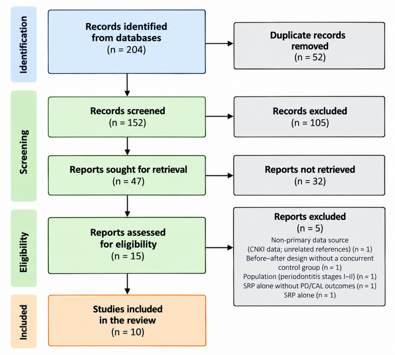
Figure 1. PRISMA 2020 flow diagram of study selection.
Note: aPDT = antimicrobial photodynamic therapy. Source: Own elaboration.Characteristics of the included studies
The ten included trials were published between 2019 and 2026 and enrolled 10 study populations totalling roughly 200 participants. Six trials were conducted in India, and one each in Italy, Iraq, Greece and Poland/Belgium. Eight used a split-mouth or site-level design, in which each participant contributed both a treated and a control site, and two were parallel-arm trials; only Annunziata 2023 and Vakaki 2025 reported a registered protocol. The photosensitizer was a phenothiazine dye, methylene blue or toluidine blue, in six trials, and indocyanine green in three, with activation wavelengths between 635 and 940 nm; one trial applied a home dual-light device. The number of sessions ranged from one to several, and follow-up ranged from three weeks to six months, with three months the most frequent assessment point. Sample sizes were small, generally between 11 and 32 participants, and the unit of analysis alternated between patients and sites, which complicates the comparison of the estimates (Table 1).
Study (year) | Design | n | Population | aPDT protocol | Follow-up |
|---|---|---|---|---|---|
Kataria 2019 | Split-mouth RCT | 30 patients (60 sites) | Chronic periodontitis, PD >= 6 mm | TBO 1%, 660 nm diode | 1 and 3 mo |
Pal 2019 | Three-arm split-mouth | 11 patients (30 sites) | Generalized periodontitis, PD > 5 mm | MB 0.3%, 940 nm diode | 3 wk |
Mallineni 2020 | Split-mouth RCT | 15 patients (30 sites) | Untreated periodontitis, PD >= 5 mm | TBO (PAD Plus), 635 nm diode | 1 and 3 mo |
Annunziata 2023 | Parallel RCT | 24 patients | Stage III grade B generalized | ICG 1 mg/ml, 810 nm diode, 2 sessions | 3 and 6 mo |
El Mobadder 2023 | Parallel RCT | 32 patients | Periodontitis, PPD > 5 mm | Tolonium chloride, 635 nm diode | 3 mo |
Najm 2024 | Split-mouth RCT | 18 patients (332 sites) | Periodontitis, PPD >= 5 mm | MB or TBO, 2 sessions | 3 mo |
Acharyya 2025 | Three-arm split-mouth | 20 patients (60 sites) | Periodontitis, PD >= 5 mm | Activated MB, 810 nm diode | 3 wk |
Vakaki 2025 | Parallel RCT | 60 smokers (25/27) | Stage III-IV in smokers | ICG dual-light, 405 + 810 nm, home daily | 4 and 6 mo |
Gupta 2026 | Three-arm site-level | 15 patients (45 sites) | Periodontitis, PPD 5-10 mm | ICG 810 nm or erythrosine LED | 15 d and 3 mo |
Lingesan 2026 | Three-arm RCT | 15 patients (60 sites) | Periodontitis, PD 5-7 mm | ICG | 4 wk |
Note: aPDT = antimicrobial photodynamic therapy; MB = methylene blue; TBO = toluidine blue O; ICG = indocyanine green; SRP = scaling and root planing; NSPT = non-surgical periodontal therapy; mo = months; wk = weeks; d = days. Source: Own elaboration.
The trials differed in the detail with which they described the procedures. Kataria 2019 used acrylic stents and a split-mouth allocation with one quadrant per treatment, but did not state whether the examiner was masked, and its report emphasized that the between-group difference in probing depth disappeared by three months.(21) Mallineni 2020 was described as triple-masked and reported full-mouth plaque, bleeding and probing measures together with a polymerase-chain-reaction count of Porphyromonas gingivalis, and its aPDT group achieved the largest separation from the control of any trial in this review.(22) Annunziata 2023 was the most rigorous: a registered, single-blind parallel trial with concealed allocation, repeated indocyanine green applications and an analysis stratified by initial pocket depth, and its overall result was null while the deep-pocket subgroup favored the therapy.(23) El Mobadder 2023 treated two parallel groups of sixteen patients with tolonium chloride and reported statistically significant advantages in both probing depth and attachment at three months.(24) Najm 2024 compared methylene blue and toluidine blue O against control sites within the same mouth and reported its primary result as the odds of pocket closure, which favored both dyes.(25) Gupta 2026 and Lingesan 2026 were small three-arm trials that used indocyanine green and reported larger probing depth reductions in the aPDT arm than in the scaling-and-root-planing-alone arm, with the caveat of five participants per group.(26,27) Pal 2019 and Acharyya 2025 measured the outcomes at three weeks only and reported their data partly in graphical form, which limited the extraction of dispersion measures.(28,29) Vakaki 2025 was the single trial conducted strictly in stage III-IV smokers and the only one whose intervention was self-administered at home, and it found no between-group difference.(30)
The classification of the included populations requires a note. Only Annunziata 2023 and Vakaki 2025 reported the stage of periodontitis according to the 2017 system; the remaining trials described severe or generalized periodontitis through probing depth and bone loss thresholds that correspond to, but do not formally document, stages III and IV. The review therefore describes the population as stage III-IV or severe periodontitis, and this approximation is treated as a limitation in the Discussion.
Risk of bias
Risk of bias was high or raised concerns in most trials, mainly because of the absence of blinding and the lack of registered protocols. Only Annunziata 2023 described allocation concealment and a pre-registered plan, and its examiner was blinded although the operator was aware of the allocation; the remaining trials did not state whether the examiner was masked, and several small trials did not describe the randomization sequence in sufficient detail to judge the risk of selection bias. Deviations from the intended interventions were generally of low concern because the treatments were delivered in one or few sessions and the follow-up was short, with little opportunity for co-interventions to differ between groups. Missing outcome data were uncommon, since attrition was minimal in most trials and the analytical units were sites rather than patients in the split-mouth studies. The main concern was the measurement of the outcome: probing is a subjective measure, and without examiner blinding the risk of detection bias is substantial, which led to a high overall judgment for the split-mouth trials that did not mask the examiner and for the open-label Vakaki 2025. Selective reporting could not be excluded in any trial without a public protocol, and the absence of prospective registration for eight of the ten trials means that outcome switching cannot be ruled out (Table 2).
Study | D1 | D2 | D3 | D4 | D5 | Overall |
|---|---|---|---|---|---|---|
Kataria 2019 | S | S | L | H | S | H |
Pal 2019 | S | S | L | H | S | H |
Mallineni 2020 | S | S | L | S | S | S |
Annunziata 2023 | L | S | L | S | L | S |
El Mobadder 2023 | S | S | L | S | S | S |
Najm 2024 | S | S | L | S | S | S |
Acharyya 2025 | S | S | L | S | S | S |
Vakaki 2025 | L | S | L | H | S | H |
Gupta 2026 | S | S | L | S | S | S |
Lingesan 2026 | S | S | L | S | S | S |
Note: D1 = randomization process; D2 = deviations from intended interventions; D3 = missing outcome data; D4 = measurement of the outcome; D5 = selection of the reported result; L = low; S = some concerns; H = high. Source: Own elaboration.
Probing depth reduction
Six trials contributed probing depth data at three months. The pooled mean difference favored adjunctive aPDT by 0.77 mm (95% CI 0.46 to 1.09; p < 0.001), with moderate heterogeneity (I2 = 64%). The per-study differences ranged from 0.23 mm in Kataria 2019 and Annunziata 2023 to 1.47 mm in Mallineni 2020, and no study favored the control group; Gupta 2026 and El Mobadder 2023 contributed intermediate estimates.(21-26) Secondary outcomes pointed in the same direction: Najm 2024 reported higher odds of pocket closure with the photosensitizers than with instrumentation alone, and Annunziata 2023 found a significantly greater reduction in initially deep pockets at six months in the aPDT group. Adverse events were absent or limited to transient discomfort in the trials that reported them (Figure 2; Table 3).
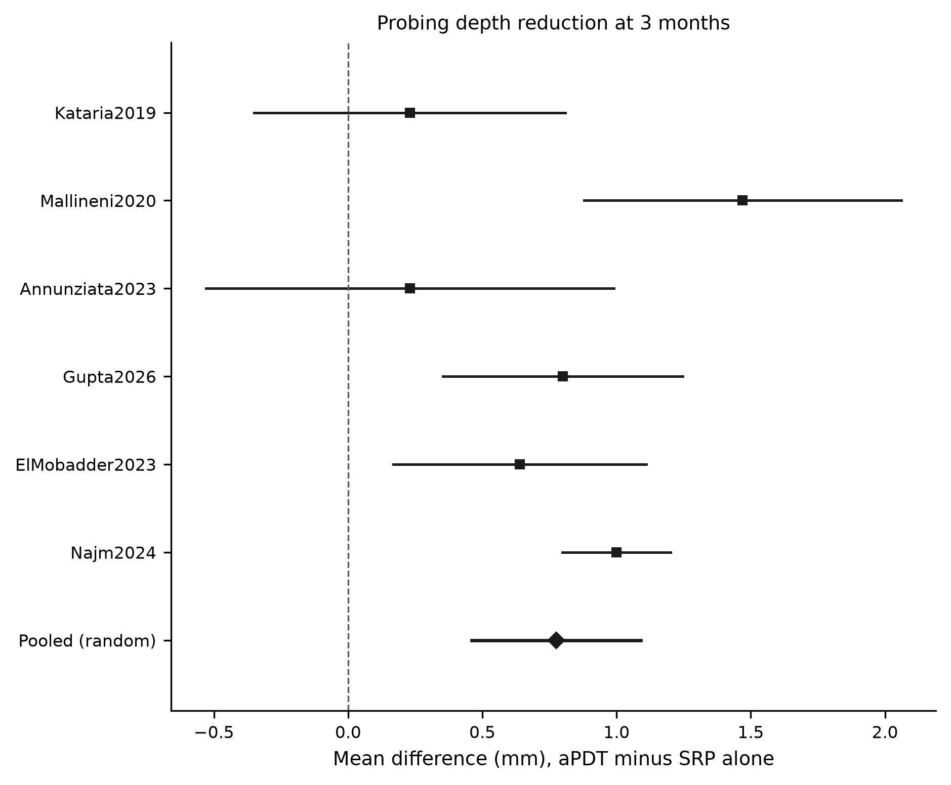
Figure 2. Forest plot of probing depth reduction at three months.
Note: Mean difference in millimetres, adjunctive aPDT minus scaling and root planing alone. Random-effects model. Source: Own elaboration.Study | n (SRP/aPDT) | SRP alone | SRP + aPDT | MD (95% CI) |
|---|---|---|---|---|
Kataria 2019 | 30 / 30 | 2.11 (1.19) | 2.34 (1.10) | 0.23 (-0.35 to 0.81) |
Mallineni 2020 | 15 / 15 | 0.73* | 2.20* | 1.47 (0.88 to 2.06) |
Annunziata 2023 | 12 / 12 | 1.54 (1.10) | 1.77 (0.77) | 0.23 (-0.53 to 0.99) |
Gupta 2026 | 15 / 30 | 0.67* | 1.47* | 0.80 (0.35 to 1.25) |
El Mobadder 2023 | 16 / 16 | 2.26* | 2.90* | 0.64 (0.17 to 1.11) |
Najm 2024 | 102 / 230 | 1.80 (0.80) | 2.80 (0.99) | 1.00 (0.80 to 1.20) |
Pooled (random) | 0.77 (0.46 to 1.09) |
Note: Change values are mean (SD) in millimetres; a positive change indicates reduction. aPDT = antimicrobial photodynamic therapy; SRP = scaling and root planing; MD = mean difference. Changes marked with an asterisk were computed from baseline and endpoint values with the SD of the change imputed (correlation 0.5). Source: Own elaboration.
Clinical attachment gain
Five trials contributed clinical attachment data at three months. The pooled mean difference was 0.72 mm in favor of adjunctive aPDT, but the confidence interval crossed zero (95% CI −0.29 to 1.74; p = 0.163) and heterogeneity was high (I2 = 89%). Four of the five point estimates favored the aPDT group, but the confidence intervals of all the individual studies except Mallineni 2020 crossed zero, so the pooled estimate does not meet a conventional threshold of significance. The largest estimate came from Mallineni 2020, in which the baseline attachment level was unbalanced between the quadrants, and Gupta 2026 was the only trial whose point estimate favored the control group (Figure 3; Table 4).
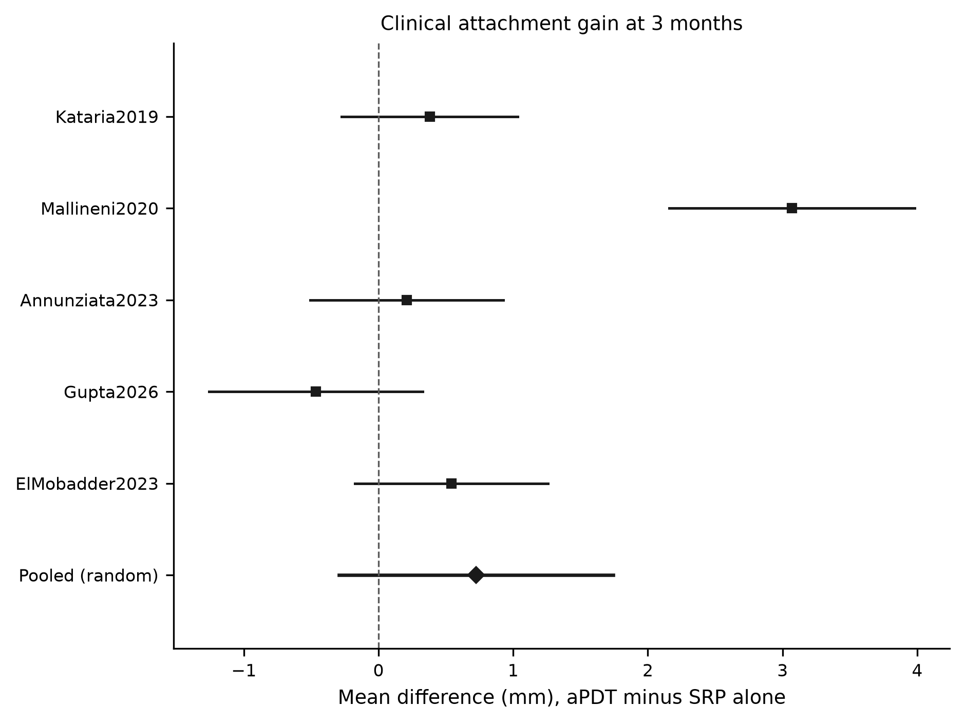
Figure 3. Forest plot of clinical attachment gain at three months.
Note: Mean difference in millimetres, adjunctive aPDT minus scaling and root planing alone. Random-effects model. Source: Own elaboration.Study | n (SRP/aPDT) | SRP alone | SRP + aPDT | MD (95% CI) |
|---|---|---|---|---|
Kataria 2019 | 30 / 30 | 1.71 (1.24) | 2.09 (1.34) | 0.38 (-0.27 to 1.03) |
Mallineni 2020 | 15 / 15 | 0.53* | 3.60* | 3.07 (2.16 to 3.98) |
Annunziata 2023 | 12 / 12 | 1.08 (0.90) | 1.29 (0.89) | 0.21 (-0.51 to 0.93) |
Gupta 2026 | 15 / 30 | 1.67* | 1.21* | -0.47 (-1.26 to 0.33) |
El Mobadder 2023 | 16 / 16 | 1.83* | 2.37* | 0.54 (-0.18 to 1.26) |
Pooled (random) | 0.72 (-0.29 to 1.74) |
Note: Change values are mean (SD) in millimetres; a positive change indicates gain. aPDT = antimicrobial photodynamic therapy; SRP = scaling and root planing; MD = mean difference. Changes marked with an asterisk were computed from baseline and endpoint values with the SD of the change imputed (correlation 0.5). Source: Own elaboration.
Sensitivity analysis
A leave-one-out analysis showed that the probing depth estimate was robust: the pooled mean difference remained between 0.67 mm and 0.88 mm and its confidence interval excluded zero whichever trial was removed. The attachment estimate was more fragile; when Mallineni 2020 was removed, the pooled mean difference fell to 0.20 mm (95% CI -0.21 to 0.60), confirming that the unbalanced trial was the main source of the apparently favorable attachment result.
Secondary outcomes
Bleeding on probing and pocket closure were reported by several trials and followed the same pattern as probing depth. Annunziata 2023 found a higher percentage of closed pockets, defined as probing depth of 4 mm or less without bleeding, in the aPDT group at six months, an advantage concentrated in initially deep pockets.(23) Najm 2024 reported that the odds of pocket closure were higher with methylene blue or toluidine blue O than with instrumentation alone, with the methylene blue protocol performing better in moderately deep pockets and the toluidine blue protocol in deep pockets.(25) Mallineni 2020 and Lingesan 2026 observed greater reductions in bleeding scores and in Porphyromonas gingivalis or total bacterial counts in the aPDT groups than in the control sites.(22,27) The microbiological changes were generally short-lived and did not always accompany the clinical improvement, which is consistent with the view that the antibacterial effect is transient and that recolonization resumes once the light-mediated decontamination ceases. Adverse events were absent or limited to transient discomfort and gingival redness in the trials that recorded them, and none of the trials reported serious complications attributable to the photodynamic therapy.
Trials with short follow-up or a home-applied device
Four trials were not included in the three-month pooling. Acharyya 2025 and Pal 2019 reported a greater probing depth reduction with activated methylene blue at three weeks than with scaling and root planing alone, but neither reached a follow-up long enough to judge clinical relevance, and both presented part of their results graphically, which precluded the extraction of standard deviations.(28,29) Lingesan 2026 found a larger mean probing depth reduction and attachment gain at four weeks with indocyanine green than with scaling and root planing alone, although the sample was small and the between-group differences in the full clinical panel did not all reach significance.(27) Vakaki 2025, the only trial restricted to stage III-IV smokers, applied a home dual-light device daily for four months and found no between-group difference in probing depth or attachment at six months, although compliance with the device was associated with better within-group outcomes and most participants reported subjective satisfaction with the device.(30) These trials suggest that any early antimicrobial advantage does not, by itself, translate into a durable clinical difference.
DISCUSSION
The pooled results indicate that adjunctive antimicrobial photodynamic therapy adds a small, statistically significant reduction in probing depth of about 0.8 mm at three months over scaling and root planing alone, but does not produce a demonstrable gain in clinical attachment. The probing depth benefit is consistent in direction across the studies, yet its magnitude is modest and sits close to the threshold usually considered clinically relevant for pocket closure. The attachment result, in turn, suggests that the extra millimetre of probing depth reduction may reflect a change in gingival position and inflammation rather than a true gain of connective tissue attachment. The clinical translation of this finding is therefore limited: a patient treated with aPDT would, on average, close a deep pocket by a fraction of a millimetre more than a patient treated with instrumentation alone, which may or may not alter the need for additional therapy.
These findings agree with earlier syntheses in the direction of effect but not in its strength. Sgolastra et al. and Atieh reported a significant additional probing depth reduction of a similar order at three months, whereas Azarpazhooh et al. found no significant difference at six months.(11-13) The divergence between the three-month and the six-month estimates in the older literature deserves attention: it suggests that any early advantage of aPDT tends to fade once the benefits of the mechanical debridement itself have matured, which is consistent with a superficial and transient antimicrobial effect rather than a regenerative one. The EFP S3 guideline, grounded on a meta-analysis of single-application protocols, concluded that the benefit did not justify a recommendation, and Salvi et al. reached the same position for untreated periodontitis.(14,16) More recent reviews restricted to indocyanine green or methylene blue protocols again describe significant but small gains in probing depth, which matches the present estimate, while acknowledging that the evidence is heterogeneous and that the clinical relevance is uncertain.(15,18,31,32) The recurrent absence of a clear attachment gain is therefore not an artifact of this review but a consistent feature of the aPDT literature, and it should temper any claim that the therapy regenerates the periodontium. In other words, the technique behaves in the literature as a decontaminating adjunct that slightly improves pocket depth in the short term, not as a regenerative or attachment-gaining intervention, and its place in therapy should be framed accordingly.
The choice of three months as the primary time point also deserves comment. Three months is the standard re-evaluation interval after subgingival instrumentation, at which the mechanical effect has largely matured, and it was the only time point available in enough trials to allow pooling. The six-month data were reported by too few of the pooled studies to be combined, but the direction of the individual results suggests that any early advantage narrows over time. This temporal pattern, together with the null six-month findings of the older reviews, supports the view that aPDT shortens the recolonization window rather than producing a lasting structural gain.( 12,14)
Individual trials outside the pooling window illustrate both sides of this literature. Braun et al., Betsy et al., Joshi et al., Hill et al., Sukumar et al., AlSarhan et al., Gandhi et al., Monzavi et al. and Moreira et al. reported superior probing depth or attachment outcomes with aPDT,(33-41) whereas Christodoulides et al., Polansky et al. and Preshaw et al. found no advantage over instrumentation alone.(42-44) The discrepant trials are instructive because they used protocols very close to those of the positive studies, and yet failed to reproduce the benefit; the null trial of Preshaw et al. is particularly relevant because it was multicentre and examiner-blinded, two design features absent from most of the positive studies.(44) This pattern raises the possibility that part of the positive signal in the smaller, unmasked trials reflects detection bias, a concern that is reinforced by the risk-of-bias assessment of the present review.
The discrepancy between studies is better explained by protocol differences than by any single factor. Photosensitizers and wavelengths vary widely, the number of sessions ranges from one to several, and some trials irradiate only the deepest sites while others treat the full mouth.(8) The concentration and pre-irradiation time of the dye, the light dose and the mode of delivery all influence the yield of reactive oxygen species, and the surfactant formulations used in some protocols were introduced precisely because the aggregation of methylene blue in aqueous solution reduces its phototoxicity. Smoking also modulates the response: trials in smokers report weaker effects, as in the home-device trial of Vakaki 2025 and in the earlier comparison of smokers and never-smokers by Theodoro et al..(30,45) The deep-pocket subgroup of Annunziata 2023, in which a significant benefit emerged only at six months, further suggests that the population and the timing of the evaluation determine whether an effect is detectable.( 23)
Heterogeneity in the present meta-analysis was moderate for probing depth and high for attachment gain. The main sources were the unit of analysis, which alternated between patients and sites, the different photosensitizers and light doses, and the baseline imbalance in attachment level in Mallineni 2020, which inflated the attachment estimate. The attachment pooling should therefore be read with caution, and the probing depth estimate, although more robust, should not be extrapolated beyond the short term. The split-mouth design, used by most of the included trials, minimizes inter-patient variability but does not eliminate the possibility of contamination between treated and control sites within the same mouth, and it complicates the estimation of the standard deviation of the change when only endpoint values are reported. A further source of divergence from other reviews is that this one admitted only photodynamic protocols, that is, those combining a photosensitizer with light, and excluded laser-only or photobiomodulation studies, whose mechanisms and dose-response differ and which are sometimes pooled together with aPDT in the broader literature.( 14)
From a mechanistic perspective, the modest clinical effect is plausible. aPDT reduces the bacterial load and inactivates lipopolysaccharide, but its action is superficial and transient, and it cannot remove the calculus or correct the anatomy that sustains the pocket; it therefore complements, rather than replaces, mechanical debridement.(7,8) This may explain why the effect is more visible in deep pockets, where instrumentation is least complete, than in shallow sites, and why repeated applications or longer wavelengths, which penetrate more deeply, are the protocols most often associated with a benefit. It also explains why the technique has not been associated with the harms of systemic antimicrobials, such as bacterial resistance or gastrointestinal effects, a property that remains its main appeal.(6,46) The absence of a corresponding attachment gain is consistent with this account: a reduction in inflammation and in pocket depth can occur through tissue shrinkage and reduced edema without the formation of new connective tissue attachment, and the latter would be required for a durable structural improvement.
This review has limitations that concern both the evidence and the review process. The number of trials is small, most were single-centre with short follow-up, and risk of bias was high or unclear in the majority; larger, registered and blinded trials are required before the technique can be recommended routinely. The populations were heterogeneous in smoking status and disease severity, and the trials that did not state the stage were classified as stage III-IV on the basis of probing depth thresholds, which may have misclassified some populations; future trials should report the stage explicitly and analyze patients, not sites, as the unit, because site-level analyses overstate precision. Only three bibliographic sources were searched, and only open-access full texts could be evaluated, so eligible trials without an accessible full text were excluded at retrieval; a broader search including the grey literature and subscription databases, together with author contact, would reduce this risk. The change standard deviations of several trials were imputed with an assumed correlation, which may distort the weights; reporting the standard deviation of the change, or the individual patient data, would remove this assumption. The three-week trials and the home-applied device were excluded from the pooling because their time point or delivery differed from the in-office protocols, and their separate description shows that the short-term effect does not guarantee a durable one. Finally, the outcome is surrogate and short-term; whether the additional probing depth reduction is maintained, and whether it translates into fewer residual pockets or less need for surgery, remains unknown and should be the target of future trials. The certainty of the evidence was not formally graded, and the small number of studies precluded the subgroup analyses, by photosensitizer or by smoking status, that would have clarified the sources of heterogeneity; a future update should register its protocol, apply GRADE and plan those subgroups in advance.
The safety profile of the intervention is favorable, and this is the main respect in which aPDT differs from systemic antibiotics. None of the included trials reported serious adverse events attributable to the therapy, and the complaints recorded were limited to transient discomfort, warmth and a sensation of excess saliva with the home device, which led to discontinuation in a minority of users.(30) This is consistent with the mechanistic premise that the phototoxic effect is confined to the sensitized and illuminated pocket and that the reactive species are short-lived.(7,8) Safety, however, does not by itself justify use, because the clinical benefit that would balance the cost and the additional chair time is still uncertain.
The comparison with established adjuncts also helps to calibrate expectations. The additional probing depth reduction observed here is of the same small order as that reported for locally delivered antimicrobials, and smaller than the gains usually attributed to systemic antimicrobials in severe disease, although the protocols, populations and time points differ enough that any direct numerical comparison would be misleading.(46,47) What distinguishes aPDT is not the size of its effect but its favorable resistance profile, which may make it attractive for patients in whom systemic antimicrobials are undesirable or contraindicated. This consideration, rather than a superiority in efficacy, is probably the most defensible basis for offering the therapy today.
The clinical meaning of the pooled probing depth difference deserves explicit comment. A mean difference of 0.77 mm is smaller than the measurement error usually attributed to manual periodontal probing, which is in the order of one millimetre, and is therefore unlikely to be perceived by the clinician in an individual patient. Its relevance depends on whether it concentrates in the deepest pockets, where a fraction of a millimetre can decide whether a pocket closes or remains patent, rather than being distributed uniformly across all sites. The subgroup data available in Annunziata 2023 and the pocket-closure analysis of Najm 2024 suggest that this may be the case, but the pooled estimate itself cannot confirm it.(23,25) An effect of this size also cannot support claims of regeneration, and the null attachment result reinforces that interpretation.
Small-study effects are a further reason for caution. The trials in this review were, with one exception, small and single-centre, and the forest plots show that the most precise estimates are those closest to zero, whereas the larger point estimates came from the smallest studies. A formal assessment of publication bias was not possible with fewer than ten studies, but the pattern is compatible with an overestimation of the effect in the small, unmasked trials, and it strengthens the case for interpreting the pooled estimate as an upper bound rather than a precise measure.
Finally, the generalizability of these findings to stage III-IV disease specifically is imperfect. Only Annunziata 2023 stated the stage explicitly among the pooled trials, and only Vakaki 2025 recruited exclusively stage III-IV patients; the remaining studies enrolled periodontitis described by probing depth thresholds that map onto, but do not prove, stages III-IV. A review that restricted itself to trials reporting the stage would have been unfeasible, but the consequence is that the pooled estimate applies to severe periodontitis broadly rather than to a verified stage III-IV population. This distinction should be borne in mind when the results are applied to individual patients.
For clinical practice, the results suggest that aPDT can be offered as an adjunct when a marginal improvement in probing depth in deep pockets is sought, but it should not replace subgingival instrumentation or be presented as an alternative to established adjuncts such as locally delivered or systemic antimicrobials, whose evidence base is more consistent.(46,47) Clinicians who elect to use it should define the photosensitizer and light parameters explicitly, treat only the sites with residual deep pockets, and re-evaluate at three and six months to judge whether the additional gain justifies the chair time and the device cost. For research, the priorities are a consensus protocol with defined light and photosensitizer parameters, examiner blinding, patient-level analyses, and outcomes that reflect pocket closure and the need for additional therapy rather than millimetre changes alone.
CONCLUSIONS
In adults with stage III-IV periodontitis, adjunctive antimicrobial photodynamic therapy produced a modest additional reduction in probing depth at three months compared with scaling and root planing alone, with moderate heterogeneity across the trials. The technique did not provide a statistically significant gain in clinical attachment, and the attachment estimate was driven by a single unbalanced trial. The available evidence is small, at moderate to high risk of bias, and therefore insufficient to support a routine recommendation in stage III-IV periodontitis. Any clinical use of the technique should be reserved for deep pockets in which a marginal additional decontamination is desired, and should be weighed against the limited and short-term nature of the benefit.
References
- 1 Tonetti MS, Jepsen S, Jin L, Otomo‐Corgel J. Impact of the global burden of periodontal diseases on health, nutrition and wellbeing of mankind: A call for global action. J Clinic Periodontology. 2017;44(5):456-462. doi: 10.1111/jcpe.12732
- 2 Tonetti MS, Greenwell H, Kornman KS. Staging and grading of periodontitis: Framework and proposal of a new classification and case definition. J Clinic Periodontology. 2018;45(S20). doi: 10.1111/jcpe.12945
- 3 Papapanou PN, Sanz M, Buduneli N, Dietrich T, Feres M, Fine DH, et al. Periodontitis: Consensus report of workgroup 2 of the 2017 World Workshop on the Classification of Periodontal and Peri‐Implant Diseases and Conditions. J Clinic Periodontology. 2018;45(S20). doi: 10.1111/jcpe.12946
- 4 Suvan J, Leira Y, Moreno Sancho FM, Graziani F, Derks J, Tomasi C. Subgingival instrumentation for treatment of periodontitis. A systematic review. J Clinic Periodontology. 2020;47(S22):155-175. doi: 10.1111/jcpe.13245
- 5 Kwon T, Lamster IB, Levin L. Current Concepts in the Management of Periodontitis. International Dental Journal. 2021;71(6):462-476. doi: 10.1111/idj.12630
- 6 Wainwright M. Photodynamic antimicrobial chemotherapy (PACT). Journal of Antimicrobial Chemotherapy. 1998;42(1):13-28. doi: 10.1093/jac/42.1.13
- 7 Soukos NS, Goodson JM. Photodynamic therapy in the control of oral biofilms. Periodontology 2000. 2011;55(1):143-166. doi: 10.1111/j.1600-0757.2010.00346.x
- 8 Cieplik F, Tabenski L, Buchalla W, Maisch T. Antimicrobial photodynamic therapy for inactivation of biofilms formed by oral key pathogens. Front Microbiol. 2014;5. doi: 10.3389/fmicb.2014.00405
- 9 Wilson M. Lethal photosensitisation of oral bacteria and its potential application in the photodynamic therapy of oral infections. Photochem Photobiol Sci. 2004;3(5):412-418. doi: 10.1039/b211266c
- 10 Nie M, Zhang P, Pathak JL, Wang X, Wu Y, Yang J, et al. Photodynamic therapy in periodontitis: A narrative review. Photoderm Photoimm Photomed. 2024;40(1):e12946. doi: 10.1111/phpp.12946
- 11 Sgolastra F, Petrucci A, Severino M, Graziani F, Gatto R, Monaco A. Adjunctive photodynamic therapy to non‐surgical treatment of chronic periodontitis: a systematic review and meta‐analysis. J Clinic Periodontology. 2013;40(5):514-526. doi: 10.1111/jcpe.12094
- 12 Azarpazhooh A, Shah PS, Tenenbaum HC, Goldberg MB. The Effect of Photodynamic Therapy for Periodontitis: A Systematic Review and Meta‐Analysis. Journal of Periodontology. 2010;81(1):4-14. doi: 10.1902/jop.2009.090285
- 13 Atieh MA. Photodynamic therapy as an adjunctive treatment for chronic periodontitis: a meta-analysis. Lasers Med Sci. 2010;25(4):605-613. doi: 10.1007/s10103-009-0744-6
- 14 Salvi GE, Stähli A, Schmidt JC, Ramseier CA, Sculean A, Walter C. Adjunctive laser or antimicrobial photodynamic therapy to non‐surgical mechanical instrumentation in patients with untreated periodontitis: A systematic review and meta‐analysis. J Clinic Periodontology. 2020;47(S22):176-198. doi: 10.1111/jcpe.13236
- 15 Dalvi S, Benedicenti S, Sălăgean T, Bordea IR, Hanna R. Effectiveness of Antimicrobial Photodynamic Therapy in the Treatment of Periodontitis: A Systematic Review and Meta-Analysis of In Vivo Human Randomized Controlled Clinical Trials. Pharmaceutics. 2021;13(6):836. doi: 10.3390/pharmaceutics13060836
- 16 Sanz M, Herrera D, Kebschull M, Chapple I, Jepsen S, Berglundh T, et al. Treatment of stage I–III periodontitis—The EFP S3 level clinical practice guideline. J Clinic Periodontology. 2020;47(S22):4-60. doi: 10.1111/jcpe.13290
- 17 Caton JG, Armitage G, Berglundh T, Chapple ILC, Jepsen S, Kornman KS, et al. A new classification scheme for periodontal and peri‐implant diseases and conditions – Introduction and key changes from the 1999 classification. Journal of Periodontology. 2018;89(S1). doi: 10.1002/jper.18-0157
- 18 Pardo A, Butera A, Giordano A, Gallo S, Pascadopoli M, Scribante A, et al. Photodynamic Therapy in Non-Surgical Treatment of Periodontitis: A Systematic Review and Meta-Analysis. Applied Sciences. 2023;13(2):1086. doi: 10.3390/app13021086
- 19 Page MJ, McKenzie JE, Bossuyt PM, Boutron I, Hoffmann TC, Mulrow CD, et al. The PRISMA 2020 statement: an updated guideline for reporting systematic reviews. BMJ. 2021:n71. doi: 10.1136/bmj.n71
- 20 Sterne JAC, Savović J, Page MJ, Elbers RG, Blencowe NS, Boutron I, et al. RoB 2: a revised tool for assessing risk of bias in randomised trials. BMJ. 2019:l4898. doi: 10.1136/bmj.l4898
- 21 Kataria P, Singh A, Shukla P, Malhotra G, Rao D. Efficacy of photodynamic therapy and lasers as an adjunct to scaling and root planing in treatment of chronic periodontitis [Internet]. 2019. Available from: https://ijmsdr.com/index.php/ijmsdr/article/view/512
- 22 Mallineni S, Nagarakanti S, Gunupati S, BV RR, Shaik MV, Chava VK. Clinical and microbiological effects of adjunctive photodynamic diode laser therapy in the treatment of chronic periodontitis: A randomized clinical trial. J Dent Res Dent Clin Dent Prospects. 2020;14(3):191-197. doi: 10.34172/joddd.2020.030
- 23 Annunziata M, Donnarumma G, Guida A, Nastri L, Persico G, Fusco A, et al. Clinical and microbiological efficacy of indocyanine green-based antimicrobial photodynamic therapy as an adjunct to non-surgical treatment of periodontitis: a randomized controlled clinical trial. Clin Oral Invest. 2023;27(5):2385-2394. doi: 10.1007/s00784-023-04875-w
- 24 El Mobadder M, Nammour S, Grzech-Leśniak K. Photodynamic Therapy with Tolonium Chloride and a Diode Laser (635 nm) in the Non-Surgical Management of Periodontitis: A Clinical Study. JCM. 2023;12(16):5270. doi: 10.3390/jcm12165270
- 25 Najm KK, Gul SS, Abdulkareem AA. Efficacy of Non-Surgical Periodontal Therapy with Adjunctive Methylene Blue and Toluidine Blue O Mediated Photodynamic in Treatment of Periodontitis: A Randomized Clinical Trial. Clinics and Practice. 2024;14(3):954-964. doi: 10.3390/clinpract14030076
- 26 Gupta P. Assessment of antimicrobial photodynamic therapy as an adjunct to scaling and root planing in periodontitis patients: A randomized controlled clinical study. Bioinformation. 2026;22(07):4116-4121. doi: 10.6026/973206300224116
- 27 L A, S R, R AA, S S, Felix JW. Comparison of Photodynamic Therapy and Laser Therapy in Conjunction with Scaling and Root Planing in Patients with Chronic Periodontitis: A Clinico-microbiological and Biochemical Study. World Journal of Dentistry. 2026;17(3):245-250. doi: 10.5005/jp-journals-10015-2776
- 28 Dept of Periodontology, Rajarajeswari Dental College and Hospital, Bangalore-560074, India, Pal DSR. Efficacy of Photodynamic Therapy and Diode laser therapy as an adjunct to Scaling and Root Planing in the treatment of Chronic Periodontitis – A Clinical & Microbiologic Short Term Study. jmscr. 2019;7(4). doi: 10.18535/jmscr/v7i4.79
- 29 Acharyya J, Raina S, Gupta R, Dahiya P, Kumar M. Effect of photodynamic therapy on non-surgical periodontal therapy in chronic periodontitis patients: a randomized clinical trial [Internet]. 2025. Available from: https://theaspd.com/index.php/ijes/article/view/4627
- 30 Vakaki C, Fragkioudakis I, Pätilä T, Sakellari D. Regular home use of dual-light photodynamic therapy as an adjunct to non-surgical periodontal treatment in smokers: a single-center randomized controlled clinical trial. Clin Oral Invest. 2025;29(12):553. doi: 10.1007/s00784-025-06600-1
- 31 Bashir NZ, Singh HA, Virdee SS. Indocyanine green–mediated antimicrobial photodynamic therapy as an adjunct to periodontal therapy: a systematic review and meta-analysis. Clin Oral Invest. 2021;25(10):5699-5710. doi: 10.1007/s00784-021-03871-2
- 32 Alasqah MN. Efficacy of methylene blue-mediated antimicrobial photodynamic therapy on clinical and radiographic outcomes among patients with periodontal diseases: A systematic review and meta-analysis of randomized controlled trials. Photodiagnosis and Photodynamic Therapy. 2024;46:104000. doi: 10.1016/j.pdpdt.2024.104000
- 33 Braun A, Dehn C, Krause F, Jepsen S. Short‐term clinical effects of adjunctive antimicrobial photodynamic therapy in periodontal treatment: a randomized clinical trial. J Clinic Periodontology. 2008;35(10):877-884. doi: 10.1111/j.1600-051x.2008.01303.x
- 34 Betsy J, Prasanth CS, Baiju KV, Prasanthila J, Subhash N. Efficacy of antimicrobial photodynamic therapy in the management of chronic periodontitis: a randomized controlled clinical trial. J Clinic Periodontology. 2014;41(6):573-581. doi: 10.1111/jcpe.12249
- 35 Joshi K, Baiju CS, Khashu H, Bansal S. Clinical effectiveness of indocyanine green mediated antimicrobial photodynamic therapy as an adjunct to scaling root planing in treatment of chronic periodontitis- A randomized controlled clinical trial. Photodiagnosis and Photodynamic Therapy. 2020;29:101591. doi: 10.1016/j.pdpdt.2019.101591
- 36 Hill G, Dehn C, Hinze AV, Frentzen M, Meister J. Indocyanine green-based adjunctive antimicrobial photodynamic therapy for treating chronic periodontitis: A randomized clinical trial. Photodiagnosis and Photodynamic Therapy. 2019;26:29-35. doi: 10.1016/j.pdpdt.2019.02.019
- 37 Sukumar K, Tadepalli A, Parthasarathy H, Ponnaiyan D. Evaluation of combined efficacy of photodynamic therapy using indocyanine green photosensitizer and non-surgical periodontal therapy on clinical and microbial parameters in the management of chronic periodontitis subjects: A randomized split-mouth design. Photodiagnosis and Photodynamic Therapy. 2020;31:101949. doi: 10.1016/j.pdpdt.2020.101949
- 38 AlSarhan MA, Altammami MA, Alaqeely RS, AlEbdi A, Jasser RA, Otaibi DA, et al. Short-term improvement of clinical parameters and microbial diversity in periodontitis patients following Indocyanine green-based antimicrobial photodynamic therapy: A randomized single-blind split-mouth cohort. Photodiagnosis and Photodynamic Therapy. 2021;35:102349. doi: 10.1016/j.pdpdt.2021.102349
- 39 Gandhi K, Pavaskar R, Cappetta E, Drew H. Effectiveness of Adjunctive Use of Low-Level Laser Therapy and Photodynamic Therapy After Scaling and Root Planing in Patients with Chronic Periodontitis. Int J Periodontics Restorative Dent. 2019;39(6):837-843. doi: 10.11607/prd.4252
- 40 Monzavi A, Chinipardaz Z, Mousavi M, Fekrazad R, Moslemi N, Azaripour A, et al. Antimicrobial photodynamic therapy using diode laser activated indocyanine green as an adjunct in the treatment of chronic periodontitis: A randomized clinical trial. Photodiagnosis and Photodynamic Therapy. 2016;14:93-97. doi: 10.1016/j.pdpdt.2016.02.007
- 41 Moreira AL, Novaes AB, Grisi MF, Taba M, Souza SL, Palioto DB, et al. Antimicrobial Photodynamic Therapy as an Adjunct to Non‐Surgical Treatment of Aggressive Periodontitis: A Split‐Mouth Randomized Controlled Trial. Journal of Periodontology. 2015;86(3):376-386. doi: 10.1902/jop.2014.140392
- 42 Christodoulides N, Nikolidakis D, Chondros P, Becker J, Schwarz F, Rössler R, et al. Photodynamic Therapy as an Adjunct to Non‐Surgical Periodontal Treatment: A Randomized, Controlled Clinical Trial. Journal of Periodontology. 2008;79(9):1638-1644. doi: 10.1902/jop.2008.070652
- 43 Polansky R, Haas M, Heschl A, Wimmer G. Clinical effectiveness of photodynamic therapy in the treatment of periodontitis. J Clinic Periodontology. 2009;36(7):575-580. doi: 10.1111/j.1600-051x.2009.01412.x
- 44 Preshaw PM, Ide M, Bissett SM, Holliday R, Lansdowne N, Pickering K, et al. No benefit of an adjunctive phototherapy protocol in treatment of periodontitis: A split‐mouth randomized controlled trial. J Clinic Periodontology. 2021;48(8):1093-1102. doi: 10.1111/jcpe.13465
- 45 Theodoro LH, Assem NZ, Longo M, Alves MLF, Duque C, Stipp RN, et al. Treatment of periodontitis in smokers with multiple sessions of antimicrobial photodynamic therapy or systemic antibiotics: A randomized clinical trial. Photodiagnosis and Photodynamic Therapy. 2018;22:217-222. doi: 10.1016/j.pdpdt.2018.04.003
- 46 Teughels W, Feres M, Oud V, Martín C, Matesanz P, Herrera D. Adjunctive effect of systemic antimicrobials in periodontitis therapy: A systematic review and meta‐analysis. J Clinic Periodontology. 2020;47(S22):257-281. doi: 10.1111/jcpe.13264
- 47 Herrera D, Matesanz P, Martín C, Oud V, Feres M, Teughels W. Adjunctive effect of locally delivered antimicrobials in periodontitis therapy: A systematic review and meta‐analysis. J Clinic Periodontology. 2020;47(S22):239-256. doi: 10.1111/jcpe.13230
Declarations
Funding
No financing
Conflict of interest
None
Authorship contributions
Drafting – original draft: Marta María Capote Fernández.
Writing–review and editing: Marta María Capote Fernández.