

Abstract
Oral manifestations were among the first extrapulmonary signs attributed to COVID-19. This narrative review traces the chronological evolution of the evidence on taste dysfunction, oral mucosal lesions and the periodontal links of COVID-19 between 2020 and 2023 and characterizes the manifestations that persist in long COVID. Searches were conducted in OpenAlex, Crossref and PubMed with an upper publication limit of 2023, and the synthesis was organized into three evidence periods and three thematic axes. In the first half of 2020, taste and smell loss emerged as frequent sentinel symptoms, pooled gustatory dysfunction reached 38.2 percent, case series described heterogeneous oral lesions and ACE2 was identified in the oral epithelium. Between mid-2020 and 2021, living systematic reviews consolidated prevalence estimates, with taste disorders in 38 percent of 64,876 patients, and case-control studies associated periodontitis with severe outcomes, including a death odds ratio of 8.81. From 2022 to 2023, persistent taste dysfunction was estimated in 4.4 percent of patients, the Omicron wave reduced chemosensory involvement, the specificity of oral lesions was questioned and Mendelian randomization did not confirm a causal periodontal link. Taste dysfunction remained the most consistent oral manifestation across the trajectory, whereas mucosal lesions and the periodontal association remained partly unresolved.
Keywords
COVID-19, post-acute COVID-19 syndrome, taste disorders, oral manifestations, periodontal diseases
INTRODUCTION
Coronavirus disease 2019 (COVID-19), caused by severe acute respiratory syndrome coronavirus 2 (SARS-CoV-2), was framed in its first weeks as a respiratory infection. Within months the clinical spectrum expanded into extrapulmonary territories, and the oral cavity became one of the earliest and most debated of them. In March 2020, clinicians began to describe sudden loss of smell and taste in patients with otherwise mild disease, a pattern that distinguished COVID-19 from influenza-like illness and from earlier coronavirus epidemics.( 1-3) The symptom was unusual in two respects: it appeared early, and it appeared in patients who would otherwise have escaped clinical attention.
Gustatory and olfactory dysfunction was quickly proposed as a screening marker. A retrospective telephone survey in Italy documented chemosensory symptoms at the onset of infection in a large proportion of the 204 patients interviewed.(4) A prospective series in Daegu, South Korea, found acute anosmia or ageusia in 15.3% of 3,191 patients in the early stage of the disease and in 15.7% of those with asymptomatic to mild illness, with higher rates in women and in younger adults.(5) These observations entered case-finding guidance because chemosensory loss accompanied mild forms that laboratory-based surveillance could miss. Patients could report the symptom without any equipment, which made it useful in dental and primary care settings. The dental relevance was direct: oral health providers examine the mouth daily and could notice chemosensory complaints long before a respiratory syndrome declared itself.
In parallel, two lines of evidence moved the oral cavity from the margin to the center of the pandemic. First, angiotensin-converting enzyme 2 (ACE2), the cellular receptor of SARS-CoV-2, was shown to be highly expressed on epithelial cells of the oral mucosa and tongue, with the protease TMPRSS2 and furin completing a plausible entry machinery.(6-8) Second, case reports began to attribute heterogeneous lesions of the oral mucosa, from ulcers to vesiculobullous changes, to the infection itself.(9-11) One stream was biological and the other clinical, and together they generated both diagnostic enthusiasm and interpretive caution.
The clinical questions condensed into three axes. The first was gustatory: whether taste loss was real, how frequent it was and whether it recovered. The second was mucosal: whether the visible lesions reported in the mouth were caused by the virus or by the circumstances of illness. The third was periodontal: whether pre-existing periodontal inflammation increased the risk of severe COVID-19 and, conversely, whether the infection worsened periodontal health. These axes matured at different speeds; a chronological reading makes that unevenness visible.
The evidence changed rapidly over the following three years. What began as anecdote matured into meta-analyses of prevalence, living systematic reviews, prospective cohorts and, finally, genetically informed causal analyses. Long COVID, recognized from 2021 onward as post-acute sequelae affecting multiple systems, added a temporal dimension that the initial reports could not anticipate.(12,13) The oral manifestations of COVID-19 are now dispersed across otolaryngology, oral medicine and periodontology, and the resulting literature is large, heterogeneous and difficult to read as a single body.
A chronological synthesis is useful because it separates findings that persisted from those that were revised and identifies the methodological and viral factors behind each shift. The objective of this narrative review is to trace the chronological evolution of the evidence on taste dysfunction, oral mucosal lesions and periodontal links of COVID-19 between 2020 and 2023 and to characterize the manifestations that persist in long COVID. It addresses three questions: how the characterization of taste dysfunction and mucosal lesions evolved, which evidence consolidated the periodontal association, and which oral manifestations endure beyond the acute infection.
METHODS
This is a narrative review reported according to the Scale for the Assessment of Narrative Review Articles (SANRA). It does not claim systematic or exhaustive coverage; the purpose was to reconstruct the trajectory of the evidence and to organize it into thematic axes and chronological periods.
Searches were conducted in OpenAlex, Crossref and PubMed with an upper publication limit of December 2023. Background literature on the biology of SARS-CoV-2 entry was allowed without a lower date limit, whereas the studies that sustain the core synthesis were restricted to the period from January 2020 to December 2023, when the pandemic evidence was produced. Terms combined variants of gustatory, olfactory and taste dysfunction; oral manifestations, mucosal lesions and oral symptoms; periodontitis, periodontal disease and COVID-19 severity; and long COVID, post-acute sequelae and persistent chemosensory dysfunction. Searches were run in English, and studies in other languages were retained when the abstract provided usable data.
Candidate records returned by each source were screened by title and abstract, and potentially relevant records were kept with their bibliographic metadata and, when available, their abstract. Studies were selected when they reported primary data or quantitative synthesis on at least one of the three axes in patients with confirmed or probable COVID-19, or when they provided mechanistic or causal evidence directly relevant to those axes. Narrative commentaries, editorials and reviews were used to contextualize the trajectory and to represent the positions held at each period. Priority was given to studies with defined samples, explicit denominators and, when available, psychophysical or laboratory confirmation rather than self-report alone. No quality scoring was applied, because the aim was to describe the evolution of the evidence rather than to rank individual studies.
The chronology was divided into three periods with explicit criteria. Period 1, from January to June 2020, corresponds to the emergence of the first case series and meta-analyses. Period 2, from July 2020 to December 2021, corresponds to the consolidation of prevalence estimates, lesion classification and the first observational evidence on the periodontal link. Period 3, from January 2022 to December 2023, corresponds to long COVID persistence, the Omicron wave and the causal and mechanistic analyses. These cutoffs follow the timing of the pandemic and the publication of the key studies rather than arbitrary calendar divisions. They are coarse by design: they mark the moments at which the dominant type of evidence changed.
The synthesis was organized into three thematic axes, taste dysfunction, oral mucosal lesions and periodontal links, and each axis was summarized in a matrix that recorded the supporting and opposing studies, the level of evidence and the period in which the finding consolidated. Prevalence figures were taken directly from the published abstracts and results of the included studies; no estimate was recomputed or pooled for this review. This structure corresponds to the SANRA dimensions of justification, selection and reasoning, and it avoids the language and flow diagrams of a systematic review, which this study is not.
DEVELOPMENT
The trajectory can be read as a progressive tightening of the evidence, from early case-based claims to population estimates and causal tests. Three periods mark the inflection points, and the timeline in Figure 1 summarizes them (Figure 1). Table 1 condenses the synthesis by thematic axis (Table 1).
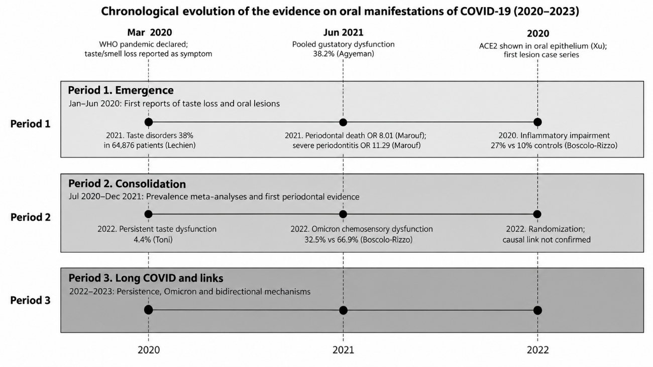
Figure 1. Chronological evolution of the evidence on oral manifestations of COVID-19, 2020-2023.
Note: Timeline of the three evidence periods with representative milestones. OR = odds ratio. Source: Own elaboration based on the reviewed literature.Thematic axis | Main findings | Period | Level of evidence |
|---|---|---|---|
Taste dysfunction | Emerged as a frequent sentinel symptom; pooled acute prevalence of 36.6-38.2%; declined during Omicron; persisted in a minority in long COVID | 2020-2023 | Moderate (meta-analyses of self-reported data; psychophysical cohorts) |
Oral mucosal lesions | Heterogeneous and uncommon; taste disturbance, xerostomia and ulcers the most frequent; direct viral causation questioned | 2020-2023 | Low to moderate (case series, cross-sectional studies, one intrahospital cohort) |
Periodontal links | Periodontitis associated with severe COVID-19 outcomes in case-control and cohort studies; causality not confirmed by Mendelian randomization | 2020-2023 | Low to moderate (observational studies with residual confounding; genetic analyses null or weak) |
Note: COVID-19 = coronavirus disease 2019. Source: Own elaboration based on the reviewed literature.
Period 1. Emergence of taste loss and the first oral lesions
The defining finding of the first months was that chemosensory dysfunction was not an incidental symptom but an early one. In an international multicenter study of adults and children, olfactory or gustatory dysfunction was present at high rates and preceded other symptoms in a substantial proportion of cases.(3) In patients presenting with influenza-like symptoms, chemosensory dysfunction was strongly associated with a positive diagnosis.(2) A telephone survey of 204 adults documented taste or smell disturbance at disease onset in most of those interviewed.(4) Because the symptom appeared in mild and even asymptomatic infections, it was proposed as an early identifier that could support testing and isolation.(1,5)
The recognition was rapid enough to matter clinically. Early commentaries argued that new-onset chemosensory loss should trigger testing and isolation precisely because it appeared in mild disease and in patients without fever, and because it preceded the respiratory phase in many cases.(1) The proposal raised a specific concern, whether smell and taste loss could be distinguished from the common cold, a question that later psychophysical work answered by showing a distinct chemosensory profile for COVID-19.(14) This early debate set the pattern for the whole literature: a plausible clinical signal, followed by a measurement problem, followed by a methodological resolution.
The public health context of the first period shaped the evidence it produced. The World Health Organization declared the pandemic in March 2020, and the early cohorts came from hospitals and outbreak investigations rather than from the community, which biased the first descriptions toward severe and hospitalized cases.( 1) The reports that followed nevertheless converged rapidly, because chemosensory loss was so noticeable to patients that it could be captured by simple questions even in telephone and online surveys.( 4,15)
The first quantitative syntheses appeared in May and June 2020. A meta-analysis of 24 studies with 8,438 test-confirmed patients estimated a pooled prevalence of gustatory dysfunction of 38.2% (95% CI 24.0 to 53.6) and of olfactory dysfunction of 41.0%.(16) A second meta-analysis of 104 studies with 38,198 patients reported substantial variation by ethnicity, with lower rates in East Asian cohorts than in European and American cohorts, a difference the authors attributed partly to reporting and partly to biology.(17) These two syntheses fixed the order of magnitude that later work would refine, and they made clear that prevalence depended on who was asked and how.
It was unclear at first whether taste loss was genuine or merely a shadow of smell loss. A position paper argued that gustatory dysfunction was specific and largely independent of olfactory dysfunction in COVID-19.( 18) A psychophysical comparison between patients with COVID-19 and patients with common colds showed that the chemosensory profile differed between the two conditions, with anosmia unaccompanied by rhinorrhoea or obstruction in COVID-19.( 14) These observations anticipated a debate that direct gustatory testing would settle later.
The same months produced the first descriptions of oral mucosal lesions attributed to the virus. A report of four patients described vesiculobullous and macular lesions and asked whether they were a true sign of the disease or coincidental.(9) Editors and clinicians debated whether oral lesions were new signs or secondary manifestations, pointing to the absence of standardized definitions and to the confounders of hospitalization.(10) A narrative synthesis catalogued the emerging descriptions, from ulcers to candidiasis and herpes-like lesions, and noted that dysgeusia was the first recognized oral symptom.(19) Reviews of this first wave agreed that the evidence was descriptive and heterogeneous.(11)
The biological plausibility of oral involvement was established in the same period. High ACE2 expression was demonstrated on epithelial cells of the oral mucosa, including the tongue,( 6) and ACE2 with furin was shown in oral epithelial cells, supporting the oral route of infection.(7,8) The presence of entry factors in human oral tissue was confirmed shortly after,( 20) and saliva was recognized as both a diagnostic fluid and a potential reservoir of the virus. Salivary gland epithelium and the tongue dorsum showed the highest signal, which placed the oral cavity on the map of susceptible tissues and motivated the use of saliva for viral detection.(8,20) On this basis, authors proposed that poor oral health and periodontitis could amplify systemic inflammation and worsen COVID-19 outcomes,(21) and a molecular analysis predicted that microRNAs upregulated by periodontitis regulate SARS-CoV-2 oral receptor genes.(22) These were hypotheses rather than clinical evidence, but they defined the periodontal question for the following years.
Period 2. Consolidation of prevalence, lesion classification and first periodontal evidence
The inflection point of the second period was the shift from case counting to population estimation. A living systematic review of oral signs and symptoms in COVID-19 summarized 40 studies with 10,228 patients in its first version(23) and, in its six-month update, 183 studies with 64,876 patients.(24) The update estimated an overall prevalence of taste disorders of 38% (95% CI 22 to 56) and reported pooled prevalences of 34% for hypogeusia, 33% for dysgeusia and 26% for ageusia.(24) The same update quantified the diagnostic value: taste disorders were associated with a positive COVID-19 test with an odds ratio of 7.54 (95% CI 5.24 to 10.86), a figure that supported their use in screening algorithms.(24) This series established that taste disturbance, not visible lesions, was the dominant oral manifestation.
Geographical variation remained the most striking feature of the prevalence estimates. A rapid systematic review of 67 studies with 27,687 confirmed cases reported taste alteration prevalences ranging from 11% in Korea to 88.8% in Belgium.(25) A meta-analysis of 138,015 patients who tested positive, drawn from papers published in 2020 and 2021, estimated that 36.6% reported taste dysfunction and concluded that direct measures of taste were at least as sensitive as self-report, which supports taste loss as a genuine symptom.(26) In children, a meta-analysis confirmed that smell or taste dysfunction occurred across pediatric ages, at lower rates than in adults.(27) Prospective surveys added the temporal dimension: in a cohort of 655 Brazilian patients, most chemosensory losses recovered within weeks, and a minority persisted at one to four months.(15)
The measurement debate was resolved in this period. Self-reported prevalence depended on the questionnaire and the population, whereas psychophysical testing produced stable and reproducible estimates and showed that gustatory impairment could exist without a parallel olfactory deficit.(18,26) This distinction mattered for clinicians, because a patient who reports taste loss is not necessarily describing the same deficit as a patient with measured gustatory threshold elevation. The living systematic review methodology itself became the vehicle of consolidation: it allowed prevalence to be updated as the denominator grew, which is why the estimate of taste disorders settled near 38% across hundreds of thousands of patients rather than drifting with each new survey.( 24)
Alongside the clinical association studies, the inflammatory hypothesis of the periodontal link was articulated in this period. Periodontitis is a microbially driven chronic inflammatory disease that raises systemic inflammatory markers, and COVID-19 severity is likewise driven by an exaggerated inflammatory response, so the two conditions were proposed to share a final common pathway of cytokine release and endothelial dysfunction.(28) The hypothesis predicted that patients with untreated periodontitis would show higher levels of systemic inflammation during infection, a prediction that later biomarker work examined directly.(29) This framing moved the debate from whether an association existed to which mediator carried it.
Recovery was the second new theme of this period. Self-reported and objective follow-up documented that taste returned within days to weeks in most patients, and that xerostomia overlapped with gustatory dysfunction in a proportion of cases.( 30) Systematic reviews consolidated the prognostic picture, showing wide but generally favorable recovery rates.(31,32) The consistent direction was that acute taste loss was self-limited in most patients, which made the persistent minority all the more relevant when long COVID emerged.
Oral mucosal lesions were progressively classified rather than merely listed. A meta-analysis of 74 studies estimated pooled prevalences of 0.33 (95% CI 0.11 to 0.60) for oral lesions, 0.44 (0.36 to 0.52) for xerostomia and 0.10 (0.01 to 0.24) for aphthous lesions, with high heterogeneity and a distinction between iatrogenic lesions and opportunistic infections.(33) Cross-sectional studies reported dry mouth in 44.2% and oral lesions in 22.4% of 204 infected patients,(34) and a series of 195 patients found oral manifestations in 29%, dominated by taste disorders (60%), xerostomia (42%) and ulcers (11%), with no association with disease severity.(35) Reviews of hospitalized patients and of the wider literature converged on the view that most lesions reflected secondary processes.(36,37)
The reassessment of oral lesions followed a consistent logic. As denominators grew, the apparent diversity of lesions narrowed into a short list, and each item on that list had an alternative explanation. Ulcers mapped onto drug reactions and mechanical injury from intubation, candidiasis onto corticosteroid use and immunosuppression, and herpes-like lesions onto viral reactivation.(36,37) This did not exclude a direct viral effect, but it shifted the burden of proof toward studies that could control for these confounders, a burden the next period would take up.
The periodontal axis moved from hypothesis to observational evidence in this period. A case-control study using the national electronic health records of Qatar, with 568 patients, found that periodontitis was associated with death (odds ratio [OR] 8.81, 95% CI 1.00 to 77.7), intensive care admission (OR 3.54, 1.39 to 9.05) and assisted ventilation (OR 4.57, 1.19 to 17.4) after adjustment for demographic, medical and behavioral factors.(38) A second case-control study reported that severe periodontitis carried an OR of 11.75 (3.89 to 35.49) for COVID-19.(39) A clinical assessment of 82 patients linked higher periodontitis severity to assisted ventilation (OR 7.45) and death (OR 14.58).(40) Retrospective cohorts in tertiary hospitals reproduced the direction of the association,(41) and the findings extended the earlier proposal that periodontal bacterial dissemination and the inflammatory state of periodontitis could favor severe lung infection in older adults.(42)
Period 3. Long COVID, Omicron and the bidirectional periodontal links
The third period was defined by the recognition of post-acute sequelae. Long COVID was established as a multisystem condition affecting a substantial minority of survivors,(12,13) and oral medicine began to ask which oral manifestations belonged to it.(43,44) Selected clinical cases documented persistent oral symptoms, including taste and salivary complaints, months after the acute episode.(45)
The definitional work of this period matters for the oral findings. Post-acute sequelae were defined as symptoms persisting beyond four weeks, and the burden was substantial, with manifestations across respiratory, neurological and other domains.(12,13) For oral health, the consequence was practical: patients with long COVID present to dental care with persistent dry mouth, taste complaints and functional limitation, and the dental team was advised to recognize these complaints and to adapt care.(43,44) A practice-oriented summary of smell and taste dysfunction after COVID-19 reinforced the need for structured follow-up of chemosensory symptoms rather than reassurance alone.(46)
For taste, the question shifted from acute prevalence to persistence. A meta-analysis with parametric cure modelling of 18 studies and 3,699 patients estimated that 4.4% (95% CI 1.2 to 14.6) of patients would have persistent taste dysfunction and 5.6% persistent smell dysfunction, with 98.0% reporting taste recovery by 180 days.(47) A matched case-control study with psychophysical testing at a median of 401 days found gustatory impairment in 27% of cases versus 10% of controls.(48) In a Brazilian survey, taste disorder was reported by 67.4% at diagnosis and still by 50% after a mean of 179 days.(49) A French cohort reported that one third of patients had gustatory dysfunction at nine months.(50) Two-year and three-year evaluations confirmed that a minority retained measurable impairment and that self-report overestimated the objective deficit.(51-53) Persistent taste and salivary sequelae were also documented in dedicated oral studies,(54,55) and a systematic review of long COVID confirmed persistent chemosensory dysfunction as a core component.(56) The mechanisms of taste injury and its proposed treatments were consolidated in reviews of the pathogenesis.(57-59)
The clinical weight of persistent taste loss extended beyond the measurement itself. Studies of long COVID reported that chemosensory dysfunction correlated with reduced quality of life and with nutritional and psychological consequences, and that the deficit had an objective basis beyond self-report.(53,55) Objective confirmation matters because the treatment options are limited and mostly supportive, which makes honest counseling about prognosis more useful than intervention without evidence.(58) The oral sequelae of the infection, dry mouth and gustatory impairment, were therefore incorporated into the post-acute assessment rather than treated as transient curiosities.(54)
The Omicron wave marked a further inflection point. In a prospective comparison, self-reported chemosensory dysfunction fell from 66.9% (95% CI 62.3 to 71.3) in the March to April 2020 cohort to 32.5% (27.6 to 37.8) in the January to February 2022 Omicron cohort, and altered taste dropped from 57.4% to 26.9%.(60) A cross-sectional survey in Shanghai during the Omicron period found chemosensory dysfunction in 44% of 686 mild or asymptomatic patients,(61) and a systematic review confirmed lower olfactory dysfunction with Omicron than with earlier variants.(62) Taste loss did not disappear, but it became less frequent and less specific as a marker, which reduced its screening value even as it remained clinically relevant.(61) Recovery after Omicron-acquired infection appeared faster and more complete than after wild-type infection, consistent with the lower initial prevalence.(62) Table 2 summarizes the trajectory of the prevalence estimates across the three periods (Table 2).
Study | Design | Sample | Prevalence of taste dysfunction (95% CI) |
|---|---|---|---|
Agyeman et al., 2020 | Systematic review and meta-analysis | 24 studies; 8,438 patients | 38.2% (24.0-53.6) |
Cirillo et al., 2021 | Rapid systematic review | 67 studies; 27,687 patients | 11.0-88.8% by country |
dos Santos et al., 2021 | Living systematic review update | 183 studies; 64,876 patients | 38.0% (22.0-56.0) |
Hannum et al., 2023 | Systematic review and meta-analysis | 2020-2021 papers; 138,015 patients | 36.6% |
Boscolo-Rizzo et al., 2022 | Prospective cohort, Omicron vs wild-type | 779 patients | 26.9% (22.3-32.0) vs 57.4% (52.6-62.0) |
dos Reis et al., 2022 | Cross-sectional survey | 305 patients | 67.4% at diagnosis; 50.0% at 179 days |
Note: CI = confidence interval. Source: Own elaboration based on the reviewed literature.
The specificity of oral mucosal lesions was actively questioned. In a prospective cohort of 154 hospitalized patients examined twice weekly, ulcerative lesions were attributed to opportunistic infections and to intubation rather than to the virus, and the authors concluded that ulcerative lesions were unlikely to be a direct manifestation of SARS-CoV-2.(63) A critical review asked whether the reported oral lesions were true manifestations or epiphenomena,(64) and a systematic reflection updated the clinical knowledge with the same caution.(65) A systematic review of 79 studies with 13,252 patients estimated pooled prevalences of 48% for taste alteration, 35% for xerostomia and 21% for ulceration, with the tongue as the most frequent site.(66) Mechanistic reviews attributed the lesions to a combination of direct viral tropism, immune dysregulation, thrombosis and secondary infection.(67) The net effect of this period was to close the lesion question in the direction of caution. By 2023, the consensus was that taste alteration and xerostomia were the reproducible oral findings, that ulcerative lesions were mostly secondary, and that the burden of proof for a specific COVID-19 lesion rested on studies with serial examination and standardized definitions.(65,66) This did not diminish the relevance of the oral examination, but it changed what the examination was expected to find.
Research on the periodontal axis acquired a bidirectional and mechanistic character. Cross-sectional studies linked oral microbiome dysbiosis to symptom severity and to the duration of COVID-19 and long COVID.(68-70) Periodontitis-derived neutrophil extracellular traps were proposed as a contributor to severe COVID-19.(71) Biomarker studies associated periodontitis severity with higher C-reactive protein and interleukin-6 in hospitalized patients,(29) and bioinformatic and immunological reviews mapped the shared pathways.(72-74) The association was also framed as bidirectional: periodontitis could aggravate the systemic response to the virus, and the infection, through endothelial damage and immune dysregulation, could in turn influence periodontal tissues.(28,74) The European Federation of Periodontology framed the association as a plausible but not fully established link.(75) The causal test arrived with Mendelian randomization: one bidirectional analysis found no causal relationship between periodontitis and hospitalization for COVID-19,(76) whereas another reported a weak positive association between COVID-19 risk and periodontitis (OR 1.02 to 1.03).(77) The genetically informed evidence therefore did not reproduce the magnitude of the observational estimates.
The oral-gut-lung axis provided a unifying narrative for the periodontal and long COVID findings. Alterations of the oral and gut microbiomes were documented during infection, and an inflammation-type dysbiosis of the oral microbiome was associated with the duration of symptoms and with the long COVID state, linking the mouth to the systemic persistence of the disease.(69,70) This axis did not prove that treating the mouth would shorten long COVID, but it identified the oral microbiome as a measurable participant in the post-acute syndrome.( 68)
DISCUSSION
The trajectory reconstructed here shows that the three oral axes of COVID-19 followed different fates. Taste dysfunction consolidated as a real, early and partly persistent symptom; oral mucosal lesions lost specificity as the evidence matured; and the periodontal link remained an association that observational studies supported but genetic analyses did not confirm.
The evolution of the taste literature is best explained by measurement and by the virus itself. Early self-reported surveys produced widely divergent estimates, from 11% to 88.8%, a spread driven by geography, case mix and the instruments used.(17,25) When direct gustatory testing was applied, the estimates stabilized and confirmed that taste loss was genuine rather than a by-product of olfactory impairment.(26) The decline observed during the Omicron wave, from 57.4% to 26.9% for altered taste, indicates that viral properties, not only methods, modulate the symptom.(60) This double dependency explains why a single prevalence figure was never adequate, and why the summary value of roughly one third masks a spread that ranges from one tenth to almost nine tenths depending on the cohort. The ethnic gradient in chemosensory dysfunction, from lower rates in East Asian cohorts to higher rates in European cohorts, was attributed to a combination of reporting norms, the viral strains circulating at the time and possibly genetic variation in ACE2.( 17) The gradient persisted in the Omicron literature, although at lower absolute rates, which argues that host and reporting factors, not the variant alone, set the baseline.( 62)
The controversy over oral mucosal lesions reflects the same problem in a more acute form. Early reviews described ulcers, candidiasis and vesiculobullous changes as oral manifestations of the disease,(19) whereas the prospective intrahospital cohort concluded that ulcerative lesions were attributable to opportunistic infection and intubation rather than to the virus.(63) The disagreement is methodological rather than contradictory: hospital cohorts that examine the mucosa serially can separate pre-existing, opportunistic and iatrogenic lesions, whereas case reports cannot.(64) The practical implication is that a visible oral lesion in a patient with COVID-19 should not be presumed specific without excluding the usual causes, and that the term oral manifestation should be reserved for findings that survive that exclusion.
Periodontal disease shows the widest gap between observational and causal evidence. Case-control studies reported large effect sizes, with odds ratios for death of 8.81 in Qatar and 14.58 in an Indian cohort,(38,40) and of 11.75 for severe periodontitis.( 39) These estimates almost certainly overstate the causal effect, because periodontitis clusters with age, diabetes and smoking, which themselves predict severe COVID-19. The Mendelian randomization studies, which are less vulnerable to that confounding, found either no causal relationship( 76) or an association of trivial magnitude.( 77) The discrepancy between an odds ratio above 8 and a genetic estimate near 1 is the clearest signal in this literature that confounding, rather than biology, explains much of the observational signal.
The mechanistic literature nonetheless provides a coherent biology. ACE2 and TMPRSS2 in the oral and lingual epithelium give SARS-CoV-2 a plausible oral entry,(6,20) and the non-neuronal expression of entry genes in the olfactory epithelium, with infection of sustentacular cells, explains how the virus disrupts chemosensation without necessarily infecting sensory neurons.(78,79) The expression data also offer a partial explanation for the demographic pattern of chemosensory loss: age- and sex-dependent expression of ACE2 and TMPRSS2 in oral epithelial cells has been reported, consistent with the higher rates of taste loss in younger patients and in women.(5,80) The original tissue-distribution maps of ACE2, published during the SARS epidemic, had already documented the receptor on the oral and lingual mucosa,(81) and the SARS-CoV-2 studies extended this anatomy to the chemosensory periphery.(82) Reviews of the mechanism of smell loss converged on injury to the supporting cells of the olfactory epithelium rather than to the sensory neurons themselves.(83,84) These mechanisms, together with the oral microbiome dysbiosis documented in patients with prolonged symptoms, connect the oral cavity to the systemic trajectory of the disease.(68,69) They do not, by themselves, establish clinical causality.
This synthesis agrees with the position taken by the European Federation of Periodontology, which described the periodontal link as plausible but not proven,(75) and with reviews that emphasized crosstalk and shared inflammatory pathways rather than a single causal direction.(28) A Spanish-language review of oral diseases associated with COVID-19 reached similar conclusions about the heterogeneity of the manifestations.(85) Where this review differs is in making the temporal dimension explicit: the same finding can be interpreted differently depending on the period in which it was produced.
The chronological design is the main strength of this review. It makes visible a pattern that a purely thematic summary would obscure: each axis was settled by a different method at a different time, taste by meta-analysis and psychophysics, mucosal lesions by prospective serial examination, and the periodontal link only partially by genetic instruments. This perspective also explains why the literature contains so many apparent contradictions; they are often statements made at different stages of the same accumulating evidence rather than genuine disagreements.
Three clinical implications follow. First, dentists and physicians should continue to treat sudden taste loss as a marker of infection, even if its sensitivity has declined with recent variants.(61) Second, patients with persistent taste complaints after COVID-19 deserve objective assessment, because self-report overestimates the deficit and because the dysfunction correlates with reduced quality of life.(53) Third, periodontal health remains a reasonable target for prevention, but it should not be presented to patients as a proven determinant of COVID-19 severity, and clinical messaging should separate the established association from the unproven causal claim. In the dental setting, the recognition of taste loss as an early marker also supported symptom screening at triage, although the decline in its sensitivity with Omicron means it cannot be the sole gatekeeper.( 61)
This review has limitations, each with a corresponding remedy. The selection was narrative and not exhaustive, so a systematic update with explicit eligibility criteria would strengthen the prevalence estimates.( 16) Searches were run mainly in English and in three bibliographic sources, which may underrepresent studies from other regions; multilingual and regional databases would widen the coverage. The heterogeneity of self-reported measures limits comparability, and a core set of psychophysical outcomes should be adopted in future cohorts. The periodontal evidence is observational and confounded, and only prospective cohorts with standardized periodontal examination can resolve the residual uncertainty. Case reports dominated the early lesion literature and are subject to publication bias, which standardized case definitions would reduce. Several prevalence figures were drawn from abstracts when the full text was not accessible, which is acceptable for orientation but not for precise meta-analysis.
The research agenda that emerges is concrete. Longitudinal psychophysical cohorts should quantify the long-term course of taste dysfunction by variant and by severity. Mendelian randomization with stronger instruments and non-European ancestries would test the causal periodontal hypothesis more decisively. Trials of periodontal treatment on COVID-19 outcomes would move the field from association to intervention, and standardized definitions of oral lesions would make the next generation of observational studies comparable.
CONCLUSIONS
Taste dysfunction was the first oral manifestation of COVID-19 to consolidate as a real symptom, and it remains the most consistent one, with pooled acute prevalences around one third and persistence in a minority of patients in long COVID. Oral mucosal lesions proved heterogeneous, uncommon and mostly secondary to opportunistic infection or to care, and their specificity was progressively revised. The periodontal link reached an association of periodontitis with severe COVID-19 outcomes in observational studies, but the causal evidence remained weak and genetically informed analyses did not confirm a large effect. Taken together, the three years of evidence separated a durable finding, taste dysfunction, from two questions that remain open: the nature of the oral lesions and the causal role of periodontitis. For clinical practice, these distinctions translate into three messages: screen for taste loss without overclaiming its specificity, examine oral lesions without presuming a viral cause, and manage periodontitis as a general health measure rather than as a proven COVID-19 modifier.
References
- 1 Xydakis MS, Dehgani-Mobaraki P, Holbrook EH, Geisthoff UW, Bauer C, Hautefort C, et al. Smell and taste dysfunction in patients with COVID-19. The Lancet Infectious Diseases. 2020;20(9):1015-1016. doi: 10.1016/s1473-3099(20)30293-0
- 2 Yan CH, Faraji F, Prajapati DP, Boone CE, DeConde AS. Association of chemosensory dysfunction and COVID‐19 in patients presenting with influenza‐like symptoms. Int Forum Allergy Rhinol. 2020;10(7):806-813. doi: 10.1002/alr.22579
- 3 Qiu C, Cui C, Hautefort C, Haehner A, Zhao J, Yao Q, et al. Olfactory and Gustatory Dysfunction as an Early Identifier of COVID‐19 in Adults and Children: An International Multicenter Study. Otolaryngol--head neck surg. 2020;163(4):714-721. doi: 10.1177/0194599820934376
- 4 Mercante G, Ferreli F, De Virgilio A, Gaino F, Di Bari M, Colombo G, et al. Prevalence of Taste and Smell Dysfunction in Coronavirus Disease 2019. JAMA Otolaryngol Head Neck Surg. 2020;146(8):723. doi: 10.1001/jamaoto.2020.1155
- 5 Lee Y, Min P, Lee S, Kim SW. Prevalence and Duration of Acute Loss of Smell or Taste in COVID-19 Patients. J Korean Med Sci. 2020;35(18):e174. doi: 10.3346/jkms.2020.35.e174
- 6 Xu H, Zhong L, Deng J, Peng J, Dan H, Zeng X, et al. High expression of ACE2 receptor of 2019-nCoV on the epithelial cells of oral mucosa. Int J Oral Sci. 2020;12(1):8. doi: 10.1038/s41368-020-0074-x
- 7 Zhong M, Lin B, Pathak JL, Gao H, Young AJ, Wang X, et al. ACE2 and Furin Expressions in Oral Epithelial Cells Possibly Facilitate COVID-19 Infection via Respiratory and Fecal–Oral Routes. Front Med. 2020;7:580796. doi: 10.3389/fmed.2020.580796
- 8 Sakaguchi W, Kubota N, Shimizu T, Saruta J, Fuchida S, Kawata A, et al. Existence of SARS-CoV-2 Entry Molecules in the Oral Cavity. IJMS. 2020;21(17):6000. doi: 10.3390/ijms21176000
- 9 Cruz Tapia RO, Peraza Labrador AJ, Guimaraes DM, Matos Valdez LH. Oral mucosal lesions in patients with SARS‐CoV‐2 infection. Report of four cases. Are they a true sign of COVID‐19 disease?. Special Care in Dentistry. 2020;40(6):555-560. doi: 10.1111/scd.12520
- 10 Amorim dos Santos J, Normando AGC, Carvalho da Silva RL, De Paula RM, Cembranel AC, Santos-Silva AR, et al. Oral mucosal lesions in a COVID-19 patient: New signs or secondary manifestations?. International Journal of Infectious Diseases. 2020;97:326-328. doi: 10.1016/j.ijid.2020.06.012
- 11 Ansari R, Gheitani M, Heidari F, Heidari F. Oral cavity lesions as a manifestation of the novel virus (COVID‐19). Oral Diseases. 2021;27(S3):771-772. doi: 10.1111/odi.13465
- 12 Parotto M, Gyöngyösi M, Howe K, Myatra SN, Ranzani O, Shankar-Hari M, et al. Post-acute sequelae of COVID-19: understanding and addressing the burden of multisystem manifestations. The Lancet Respiratory Medicine. 2023;11(8):739-754. doi: 10.1016/s2213-2600(23)00239-4
- 13 Perumal R, Shunmugam L, Naidoo K, Abdool Karim SS, Wilkins D, Garzino-Demo A, et al. Long COVID: a review and proposed visualization of the complexity of long COVID. Front Immunol. 2023;14:1117464. doi: 10.3389/fimmu.2023.1117464
- 14 Huart C, Philpott C, Konstantinidis I, Altundag A, Whitcroft KL, Trecca EMC, et al. Comparison of COVID-19 and common cold chemosensory dysfunction. Rhin. 2020;58(6):623-625. doi: 10.4193/rhin20.251
- 15 Brandão Neto D, Fornazieri MA, Dib C, Di Francesco RC, Doty RL, Voegels RL, et al. Chemosensory Dysfunction in COVID‐19: Prevalences, Recovery Rates, and Clinical Associations on a Large Brazilian Sample. Otolaryngol--head neck surg. 2021;164(3):512-518. doi: 10.1177/0194599820954825
- 16 Agyeman AA, Chin KL, Landersdorfer CB, Liew D, Ofori-Asenso R. Smell and Taste Dysfunction in Patients With COVID-19: A Systematic Review and Meta-analysis. Mayo Clinic Proceedings. 2020;95(8):1621-1631. doi: 10.1016/j.mayocp.2020.05.030
- 17 von Bartheld CS, Hagen MM, Butowt R. Prevalence of Chemosensory Dysfunction in COVID-19 Patients: A Systematic Review and Meta-analysis Reveals Significant Ethnic Differences. ACS Chem Neurosci. 2020;11(19):2944-2961. doi: 10.1021/acschemneuro.0c00460
- 18 Vaira LA, Lechien JR, Salzano G, Salzano FA, Maglitto F, Saussez S, et al. Gustatory Dysfunction: A Highly Specific and Smell-Independent Symptom of COVID-19. Indian J Otolaryngol Head Neck Surg. 2022;74(S2):2755-2757. doi: 10.1007/s12070-020-02182-4
- 19 Iranmanesh B, Khalili M, Amiri R, Zartab H, Aflatoonian M. Oral manifestations of COVID ‐19 disease: A review article. Dermatologic Therapy. 2021;34(1):e14578-e14578. doi: 10.1111/dth.14578
- 20 Sawa Y, Ibaragi S, Okui T, Yamashita J, Ikebe T, Harada H. Expression of SARS‐CoV‐2 entry factors in human oral tissue. Journal of Anatomy. 2021;238(6):1341-1354. doi: 10.1111/joa.13391
- 21 Botros N, Iyer P, Ojcius DM. Is there an association between oral health and severity of COVID-19 complications?. Biomedical Journal. 2020;43(4):325-327. doi: 10.1016/j.bj.2020.05.016
- 22 Roganović JR. microRNA‐146a and ‐155, upregulated by periodontitis and type 2 diabetes in oral fluids, are predicted to regulate SARS‐CoV‐2 oral receptor genes. Journal of Periodontology. 2021;92(7):35-43. doi: 10.1002/jper.20-0623
- 23 Amorim dos Santos J, Normando AGC, Carvalho da Silva RL, Acevedo AC, De Luca Canto G, Sugaya N, et al. Oral Manifestations in Patients with COVID-19: A Living Systematic Review. J Dent Res. 2021;100(2):141-154. doi: 10.1177/0022034520957289
- 24 Amorim dos Santos J, Normando AGC, Carvalho da Silva RL, Acevedo AC, De Luca Canto G, Sugaya N, et al. Oral Manifestations in Patients with COVID-19: A 6-Month Update. J Dent Res. 2021;100(12):1321-1329. doi: 10.1177/00220345211029637
- 25 Cirillo N, Bizzoca ME, Lo Muzio E, Cazzolla AP, Lo Muzio L. Gustatory dysfunction in COVID-19 patients: a rapid systematic review on 27,687 cases. Acta Odontologica Scandinavica. 2021;79(6):418-425. doi: 10.1080/00016357.2020.1869828
- 26 Hannum ME, Koch RJ, Ramirez VA, Marks SS, Toskala AK, Herriman RD, et al. Taste loss as a distinct symptom of COVID-19: a systematic review and meta-analysis. Chemical Senses. 2023;48:bjad043. doi: 10.1093/chemse/bjad043
- 27 Yan Q, Qiu D, Liu X, Guo X, Hu Y. Prevalence of Smell or Taste Dysfunction Among Children With COVID-19 Infection: A Systematic Review and Meta-Analysis. Front Pediatr. 2021;9:686600. doi: 10.3389/fped.2021.686600
- 28 Brandini DA, Takamiya AS, Thakkar P, Schaller S, Rahat R, Naqvi AR. Covid‐19 and oral diseases: Crosstalk, synergy or association?. Reviews in Medical Virology. 2021;31(6):e2226. doi: 10.1002/rmv.2226
- 29 Moradi Haghgoo J, Torkzaban P, Farhadian M, Moosavi Sedeh SA. Association between the severity of periodontitis, COVID-19, C-reactive protein and interleukin-6 levels in hospitalized patients: a case‒control study. BMC Oral Health. 2023;23(1):556. doi: 10.1186/s12903-023-03270-x
- 30 Fantozzi PJ, Pampena E, Di Vanna D, Pellegrino E, Corbi D, Mammucari S, et al. Xerostomia, gustatory and olfactory dysfunctions in patients with COVID-19. American Journal of Otolaryngology. 2020;41(6):102721. doi: 10.1016/j.amjoto.2020.102721
- 31 Costa KVTD, Carnaúba ATL, Rocha KW, Andrade KCLD, Ferreira SMS, Menezes PDL. Olfactory and taste disorders in COVID-19: a systematic review. Brazilian Journal of Otorhinolaryngology. 2020;86(6):781-792. doi: 10.1016/j.bjorl.2020.05.008
- 32 Ahmad S, Sohail A, Shahid Chishti MA, Aemaz Ur Rehman M, Farooq H. How common are taste and smell abnormalities in COVID-19? A systematic review and meta-analysis. Journal of Taibah University Medical Sciences. 2022;17(2):174-185. doi: 10.1016/j.jtumed.2021.10.009
- 33 Aragoneses J, Suárez A, Algar J, Rodríguez C, López-Valverde N, Aragoneses JM. Oral Manifestations of COVID-19: Updated Systematic Review With Meta-Analysis. Front Med. 2021;8:726753. doi: 10.3389/fmed.2021.726753
- 34 Tuter G, Yerebakan M, Celik B, Kara G. Oral manifestations in SARS-CoV-2 infection. Med Oral. 2022;27(4):e330-e339. doi: 10.4317/medoral.25259
- 35 Binmadi NO, Aljohani S, Alsharif MT, Almazrooa SA, Sindi AM. Oral Manifestations of COVID-19: A Cross-Sectional Study of Their Prevalence and Association with Disease Severity. JCM. 2022;11(15):4461. doi: 10.3390/jcm11154461
- 36 Bhujel N, Zaheer K, Singh RP. Oral mucosal lesions in patients with COVID-19: a systematic review. British Journal of Oral and Maxillofacial Surgery. 2021;59(9):1024-1030. doi: 10.1016/j.bjoms.2021.06.011
- 37 SURBOYO MD, ERNAWATI DS, BUDI HS. Oral mucosal lesions and oral symptoms of the SARS-CoV-2 infection. Minerva Dent Oral Sc. 2021;70(4):161-168. doi: 10.23736/s2724-6329.21.04493-9
- 38 Marouf N, Cai W, Said KN, Daas H, Diab H, Chinta VR, et al. Association between periodontitis and severity of COVID‐19 infection: A case–control study. J Clinic Periodontology. 2021;48(4):483-491. doi: 10.1111/jcpe.13435
- 39 Anand PS, Jadhav P, Kamath KP, Kumar SR, Vijayalaxmi S, Anil S. A case‐control study on the association between periodontitis and coronavirus disease (COVID‐19). Journal of Periodontology. 2022;93(4):584-590. doi: 10.1002/jper.21-0272
- 40 Gupta S, Mohindra R, Singla M, Khera S, Sahni V, Kanta P, et al. The clinical association between Periodontitis and COVID-19. Clin Oral Invest. 2022;26(2):1361-1374. doi: 10.1007/s00784-021-04111-3
- 41 Alnomay N, Alolayan L, Aljohani R, Almashouf R, Alharbi G. Association between periodontitis and COVID-19 severity in a tertiary hospital: A retrospective cohort study. The Saudi Dental Journal. 2022;34(7):623-628. doi: 10.1016/j.sdentj.2022.07.001
- 42 Aquino-Martinez R, Hernández-Vigueras S. Severe COVID-19 Lung Infection in Older People and Periodontitis. JCM. 2021;10(2):279. doi: 10.3390/jcm10020279
- 43 Patton LL. Long-COVID and the practice of oral medicine. Oral Surgery, Oral Medicine, Oral Pathology and Oral Radiology. 2022;133(2):125-128. doi: 10.1016/j.oooo.2021.10.025
- 44 France K, Glick M. Long COVID and oral health care considerations. The Journal of the American Dental Association. 2022;153(2):167-174. doi: 10.1016/j.adaj.2021.08.007
- 45 Rafałowicz B, Wagner L, Rafałowicz J. Long COVID Oral Cavity Symptoms Based on Selected Clinical Cases. Eur J Dent. 2022;16(02):458-463. doi: 10.1055/s-0041-1739445
- 46 Boscolo-Rizzo P, Polesel J, Vaira LA. Smell and taste dysfunction after covid-19. BMJ. 2022;378:o1653. doi: 10.1136/bmj.o1653
- 47 Tan BKJ, Han R, Zhao JJ, Tan NKW, Quah ESH, Tan CJW, et al. Prognosis and persistence of smell and taste dysfunction in patients with covid-19: meta-analysis with parametric cure modelling of recovery curves. BMJ. 2022;378:e069503. doi: 10.1136/bmj-2021-069503
- 48 Boscolo-Rizzo P, Hummel T, Hopkins C, Dibattista M, Menini A, Spinato G, et al. High prevalence of long-term olfactory, gustatory, and chemesthesis dysfunction in post-COVID-19 patients: a matched case-control study with one-year follow-up using a comprehensive psychophysical evaluation. Rhin. 2021;0(0):0-0. doi: 10.4193/rhin21.249
- 49 Reis D, Sartoretto SC, Calasans‐Maia MD, Louro RS, Moraschini V. Long‐term prevalence of taste and olfactory dysfunction in COVID‐19 patients: A cross‐sectional study. Oral Diseases. 2022;28(S2):2516-2521. doi: 10.1111/odi.14231
- 50 Klopfenstein T, Tipirdamaz C, Gendrin V, Osman M, Mercier J, Bouvier E, et al. Third of patients have gustatory dysfunction 9 months after SARS-CoV-2 infection: the ANOSVID study. International Journal of Infectious Diseases. 2022;119:114-116. doi: 10.1016/j.ijid.2022.03.053
- 51 Boscolo-Rizzo P, Tofanelli M, Zanelli E, Gardenal N, Tirelli G. COVID-19-Related Quantitative and Qualitative Olfactory and Gustatory Dysfunction: Long-Term Prevalence and Recovery Rate. ORL. 2023;85(2):67-71. doi: 10.1159/000525861
- 52 Boscolo‐Rizzo P, Hummel T, Invitto S, Spinato G, Tomasoni M, Emanuelli E, et al. Psychophysical assessment of olfactory and gustatory function in post‐mild COVID‐19 patients: A matched case‐control study with 2‐year follow‐up. Int Forum Allergy Rhinol. 2023;13(10):1864-1875. doi: 10.1002/alr.23148
- 53 Boscolo-Rizzo P, Spinato G, Hopkins C, Marzolino R, Cavicchia A, Zucchini S, et al. Evaluating long-term smell or taste dysfunction in mildly symptomatic COVID-19 patients: a 3-year follow-up study. Eur Arch Otorhinolaryngol. 2023;280(12):5625-5630. doi: 10.1007/s00405-023-08227-y
- 54 Tsuchiya H. COVID-19 Oral Sequelae: Persistent Gustatory and Saliva Secretory Dysfunctions after Recovery from COVID-19. Med Princ Pract. 2023;32(3):166-177. doi: 10.1159/000531373
- 55 Cardoso Soares P, Moreira de Freitas P, de Paula Eduardo C, Hiramatsu Azevedo L. COVID-19-Related Long-Term Taste Impairment: Symptom Length, Related Taste, Smell Disturbances, and Sample Characteristics. Cureus. 2023;15(4):e38055-e38055. doi: 10.7759/cureus.38055
- 56 De Luca P, Di Stadio A, Colacurcio V, Marra P, Scarpa A, Ricciardiello F, et al. Long COVID, audiovestibular symptoms and persistent chemosensory dysfunction: a systematic review of the current evidence. Acta Otorhinolaryngol Ital. 2022;42(2 (Suppl.1)):S87-S93. doi: 10.14639/0392-100x-suppl.1-42-2022-10
- 57 Okada Y, Yoshimura K, Toya S, Tsuchimochi M. Pathogenesis of taste impairment and salivary dysfunction in COVID-19 patients. Japanese Dental Science Review. 2021;57:111-122. doi: 10.1016/j.jdsr.2021.07.001
- 58 Wang J, Liu R, Ma H, Zhang W. The Pathogenesis of COVID-19–Related Taste Disorder and Treatments. J Dent Res. 2023;102(11):1191-1198. doi: 10.1177/00220345231182926
- 59 Srinivasan M. Taste Dysfunction and Long COVID-19. Front Cell Infect Microbiol. 2021;11:716563. doi: 10.3389/fcimb.2021.716563
- 60 Boscolo‐Rizzo P, Tirelli G, Meloni P, Hopkins C, Madeddu G, De Vito A, et al. Coronavirus disease 2019 (COVID‐19)–related smell and taste impairment with widespread diffusion of severe acute respiratory syndrome–coronavirus‐2 (SARS‐CoV‐2) Omicron variant. Int Forum Allergy Rhinol. 2022;12(10):1273-1281. doi: 10.1002/alr.22995
- 61 Wang J, Chen Y, Huang J, Niu C, Zhang P, Yuan K, et al. Prevalence of taste and smell dysfunction in mild and asymptomatic COVID-19 patients during Omicron prevalent period in Shanghai, China: a cross-sectional survey study. BMJ Open. 2023;13(3):e067065. doi: 10.1136/bmjopen-2022-067065
- 62 von Bartheld CS, Wang L. Prevalence of Olfactory Dysfunction with the Omicron Variant of SARS-CoV-2: A Systematic Review and Meta-Analysis. Cells. 2023;12(3):430. doi: 10.3390/cells12030430
- 63 Schwab G, Palmieri M, Zerbinati RM, Sarmento DJS, Reis T, Ortega KL, et al. Lack of direct association between oral mucosal lesions and SARS-CoV- 2 in a cohort of patients hospitalised with COVID-19. Journal of Oral Microbiology. 2022;14(1):2047491. doi: 10.1080/20002297.2022.2047491
- 64 Sarasati A, Agustina D, Surboyo MDC. The Oral Lesion in the COVID-19 Patient: Is It True Oral Manifestation or Not?. IDR. 2023;Volume 16:4357-4385. doi: 10.2147/idr.s411615
- 65 Kot WY, Li JW, Chan AKY, Zheng LW. A reflection on COVID-19 and oral mucosal lesion: a systematic review. Front Oral Health. 2023;4:1322458. doi: 10.3389/froh.2023.1322458
- 66 Gupta A, Shrivastav K, Agrawal A, Purohit A, Chanchlani R. Estimating the prevalence of oral manifestations in COVID-19 patients: a systematic review. PHRP. 2023;14(5):388-417. doi: 10.24171/j.phrp.2023.0033
- 67 Lin W, Gao F, Wang X, Qin N, Chen X, Tam KY, et al. The oral manifestations and related mechanisms of COVID-19 caused by SARS-CoV-2 infection. Front Cell Neurosci. 2023;16:1006977. doi: 10.3389/fncel.2022.1006977
- 68 Soffritti I, D’Accolti M, Fabbri C, Passaro A, Manfredini R, Zuliani G, et al. Oral Microbiome Dysbiosis Is Associated With Symptoms Severity and Local Immune/Inflammatory Response in COVID-19 Patients: A Cross-Sectional Study. Front Microbiol. 2021;12:687513. doi: 10.3389/fmicb.2021.687513
- 69 Haran JP, Bradley E, Zeamer AL, Cincotta L, Salive MC, Dutta P, et al. Inflammation-type dysbiosis of the oral microbiome associates with the duration of COVID-19 symptoms and long COVID. JCI Insight. 2021;6(20):e152346. doi: 10.1172/jci.insight.152346
- 70 Ren Z, Wang H, Cui G, Lu H, Wang L, Luo H, et al. Alterations in the human oral and gut microbiomes and lipidomics in COVID-19. Gut. 2021;70(7):1253-1265. doi: 10.1136/gutjnl-2020-323826
- 71 Vitkov L, Knopf J, Krunić J, Schauer C, Schoen J, Minnich B, et al. Periodontitis-Derived Dark-NETs in Severe Covid-19. Front Immunol. 2022;13:872695. doi: 10.3389/fimmu.2022.872695
- 72 Zhang C, Sun Y, Xu M, Shu C, Yue Z, Hou J, et al. Potential links between COVID-19 and periodontitis: a bioinformatic analysis based on GEO datasets. BMC Oral Health. 2022;22(1):520. doi: 10.1186/s12903-022-02435-4
- 73 Qi M, Sun W, Wang K, Li W, Lin J, Gong J, et al. Periodontitis and COVID-19: Immunological Characteristics, Related Pathways, and Association. IJMS. 2023;24(3):3012. doi: 10.3390/ijms24033012
- 74 Brock M, Bahammam S, Sima C. The Relationships Among Periodontitis, Pneumonia and COVID-19. Front Oral Health. 2022;2:801815. doi: 10.3389/froh.2021.801815
- 75 Herrera D, Serrano J, Roldán S, Alonso B, Sanz M. Oral and systemic health: is there a “new” link with COVID-19?. Clin Oral Invest. 2023;27(S1):3-13. doi: 10.1007/s00784-023-04950-2
- 76 Song J, Wu Y, Yin X, Zhang J. Relationship between periodontitis and COVID‐19: A bidirectional two‐sample Mendelian randomization study. Health Science Reports. 2023;6(8):e1413. doi: 10.1002/hsr2.1413
- 77 Meng Z, Ma Y, Li W, Deng X. Association between periodontitis and COVID-19 infection: a two-sample Mendelian randomization study. PeerJ. 2023;11:e14595. doi: 10.7717/peerj.14595
- 78 Brann DH, Tsukahara T, Weinreb C, Lipovsek M, Van den Berge K, Gong B, et al. Non-neuronal expression of SARS-CoV-2 entry genes in the olfactory system suggests mechanisms underlying COVID-19-associated anosmia. Sci Adv. 2020;6(31):eabc5801. doi: 10.1126/sciadv.abc5801
- 79 Verma AK, Zheng J, Meyerholz DK, Perlman S. SARS-CoV-2 infection of sustentacular cells disrupts olfactory signaling pathways. JCI Insight. 2022;7(24):e160277. doi: 10.1172/jci.insight.160277
- 80 Peng J, Sun J, Zhao J, Deng X, Guo F, Chen L. Age and gender differences in ACE2 and TMPRSS2 expressions in oral epithelial cells. J Transl Med. 2021;19(1):358. doi: 10.1186/s12967-021-03037-4
- 81 Hamming I, Timens W, Bulthuis M, Lely A, Navis G, van Goor H. Tissue distribution of ACE2 protein, the functional receptor for SARS coronavirus. A first step in understanding SARS pathogenesis. The Journal of Pathology. 2004;203(2):631-637. doi: 10.1002/path.1570
- 82 Fodoulian L, Tuberosa J, Rossier D, Boillat M, Kan C, Pauli V, et al. SARS-CoV-2 Receptors and Entry Genes Are Expressed in the Human Olfactory Neuroepithelium and Brain. iScience. 2020;23(12):101839. doi: 10.1016/j.isci.2020.101839
- 83 Doty RL. The mechanisms of smell loss after SARS-CoV-2 infection. The Lancet Neurology. 2021;20(9):693-695. doi: 10.1016/s1474-4422(21)00202-7
- 84 Kanjanaumporn J, Aeumjaturapat S, Snidvongs K, Seresirikachorn K, Chusakul S. Smell and taste dysfunction in patients with SARS-CoV-2 infection: A review of epidemiology, pathogenesis, prognosis, and treatment options. Asian Pac J Allergy Immunol. 2020;38(2):69-77. doi: 10.12932/ap-030520-0826
- 85 Blanco Romero L, Figueredo Rigores A, Ortiz Figueroa AM. Oral diseases associated with COVID-19. Nursing Depths Series. 2022;1:29. doi: 10.56294/nds202229
Declarations
Funding
No financing
Conflict of interest
None
Authorship contributions
Drafting – original draft: Ana Rodríguez Yanes.
Writing–review and editing: Ana Rodríguez Yanes.