

Abstract
Introduction: Postoperative pain and periapical healing can be influenced by the root canal sealer, and bioceramic (calcium silicate-based) sealers are increasingly recommended over epoxy resin sealers despite contradictory clinical evidence. Methods: A systematic review was conducted following PRISMA 2020. PubMed, OpenAlex and Crossref were searched up to 2023 for clinical studies comparing calcium silicate-based and epoxy resin sealers in permanent teeth. Risk of bias was assessed, and random-effects meta-analyses pooled the standardized mean difference for pain at 24 hours and the risk ratio for periapical healing. Results: From 490 records, 10 studies were included (seven randomized trials and three observational studies). Individual studies mostly found no significant difference in pain; the pooled standardized mean difference at 24 hours was -0.36 (95% CI -0.66 to -0.05; p = 0.021; I² = 2.3%), favoring bioceramic sealers. Periapical healing was similar (pooled risk ratio 1.04; 95% CI 0.93 to 1.16; p = 0.497). Conclusions: Bioceramic sealers are associated with slightly lower 24-hour postoperative pain but similar periapical healing compared with epoxy resin sealers; the certainty of the evidence is low.
Keywords
bioceramic sealer, calcium silicate, epoxy resin sealer, postoperative pain, periapical healing, root canal treatment
INTRODUCTION
Endodontic postoperative pain is one of the most common and least predictable complications of root canal treatment. Its reported incidence ranges from 3% to 58% depending on the population studied, the instruments used and the definition of pain,( 1) and it is consistently associated with preoperative pain, symptomatic pulp status, apical enlargement and the degree of periapical inflammation.(2,3) The obturation step introduces one further variable. The sealer is placed in contact with the canal wall and can pass through the apical foramen, lateral canals and dentinal tubules into the periapical tissues, where its components may sustain the inflammatory response that underlies pain and may retard the repair of the periapical lesion. Because a sealer is used in virtually every obturation, small differences between materials can accumulate into clinically relevant differences in postoperative discomfort and in the probability of radiographic healing. Pain also drives analgesic consumption, unplanned visits and patient anxiety about the procedure, which makes its prevention a practical priority beyond the immediate discomfort.
Two families of sealers dominate current practice and represent different philosophies. Epoxy resin sealers, of which AH Plus is the reference, polymerize by an epoxy-amine reaction into a dense, dimensionally stable mass with high bond strength and radiopacity, and they have long been regarded as the clinical standard against which other sealers are measured.(4) Their principal disadvantage is biological: they release residual monomers, amine compounds and formaldehyde in the first hours after placement, and these leachables are cytotoxic and genotoxic to periapical cells.(5) Calcium silicate-based sealers, marketed as bioceramic or hydraulic sealers, were introduced to solve this problem. They set by hydration, release calcium and hydroxyl ions, raise the local pH and precipitate hydroxyapatite on their surface, properties that in vitro favour cell viability, mineralization and an alkaline antibacterial environment.(6,7) The clinical appeal of the bioceramic class rests on this bioactivity and on the claim that it reduces postoperative inflammation and accelerates periapical repair. The biological pathway is well characterized in vitro: freshly mixed epoxy resin sealers release amines and monomers that activate transient receptor potential and calcitonin gene-related peptide signalling in trigeminal nociceptors, and the reactive oxygen species generated by residual monomers correlate with inflammatory pain, whereas calcium silicate sealers produce far less chemical irritation and, once hydrated, become alkaline and deposit apatite.(5,8) For healing, the calcium and hydroxyl ions released by hydraulic sealers stimulate osteogenic differentiation and inhibit osteoclast activity in culture.(7,8) These mechanisms predict less early pain and faster repair with the bioceramic class, but they come from cell and animal models, and their magnitude in patients is unknown.
Whether that claim holds in patients is the unresolved question this review addresses. Individual trials have produced contradictory results. Some found no difference in postoperative pain between the two sealer families,(9,10) whereas pooled estimates have reported lower pain with bioceramic sealers at 24 and 48 hours.(11,12) The systematic reviews published on the question disagree in the same way: one concluded that calcium silicate-based sealers do not reduce the risk or intensity of postoperative pain when compared with epoxy resin sealers,(13) another reported a small advantage for bioceramic sealers at early time points,(14) and a third found comparable or slightly better healing with the calcium silicate materials.(15) The clinician therefore receives opposing messages about a decision that is made at every obturation; the disagreement is rarely explained beyond a generic call for more trials. Postoperative pain, although distressing, is transient and resolves within days in most patients; the outcome that determines the long-term fate of the tooth is the resolution of the periapical lesion. Periapical healing depends on the elimination of the intracanal infection and on a seal that prevents reinfection, and the sealer participates in both. Because healing is slow, differences between sealers are expected to appear only after months and to be smaller than the differences produced by canal preparation and disinfection, which is why pain and healing are frequently reported together and why the two outcomes can move in opposite directions in the same patient. A further complication is that the sealer is almost never changed in isolation in clinical studies: bioceramic sealers are typically placed with a single cone or a sealer-based technique, whereas epoxy resin sealers are placed with warm vertical compaction, so the comparison is between two obturation systems and not only between two chemistries.
Previous syntheses are also limited in scope. Most examined either pain( 12,14,16) or radiographic healing( 17) in isolation, pooled randomized trials without observational evidence, mixed non-epoxy comparators such as zinc oxide-eugenol or calcium hydroxide sealers into the control arm, or ended their searches before the most recent trials were published. The most recent of these reviews searched up to 2020 or 2021,(13,14) and the largest pooled nine randomized trials for pain.( 11) A review published in 2023 combined pain with radiographic healing but described the overall evidence as modest.( 18) None quantified both outcomes jointly across randomized and observational designs within a single bibliographic window, which is precisely the comparison a clinician needs when choosing between the two sealer families.
The objective of this systematic review was therefore to compare the effect of calcium silicate-based (bioceramic) sealers versus epoxy resin sealers on postoperative pain and periapical healing after root canal treatment of permanent teeth, using the question: in permanent teeth receiving root canal treatment, do bioceramic sealers reduce postoperative pain and improve periapical healing compared with epoxy resin sealers? A meta-analysis was performed where the outcome measures of the included studies were compatible.
METHODS
The review was conducted and reported according to the PRISMA 2020 statement.
The PICOS framework defined eligibility. Population: permanent teeth of adult patients receiving primary root canal treatment or non-surgical retreatment. Intervention: obturation with a calcium silicate-based (bioceramic) sealer, including premixed injectable formulations and MTA-containing pastes. Comparison: obturation with an epoxy resin sealer (AH Plus or an equivalent epoxy-amine product). Outcomes: postoperative pain, measured as incidence, intensity or analgesic intake, and periapical healing, measured as radiographic success, complete or partial healing, or reduction of the radiolucent lesion. Study design: randomized clinical trials and prospective or retrospective observational clinical studies. Records published between 2015 and 2023 in English or Spanish were eligible. Laboratory studies, animal studies, case reports, and studies comparing the bioceramic sealer with a non-epoxy control were excluded. The epoxy resin restriction was applied strictly because the review question contrasts the two chemistries, and because zinc oxide-eugenol and calcium hydroxide sealers, although commonly used as controls, behave differently and would dilute the comparison.
PubMed, OpenAlex and Crossref were searched, restricted to records published up to 2023 to respect the bibliographic window of the target volume. The query set covered three needs: core queries for the eligible clinical studies, which combined terms for the sealer families with terms for the outcomes and the procedure; product queries, which combined the commercial names of the sealers (iRoot SP, EndoSequence BC Sealer, BioRoot RCS, Endoseal MTA, Sealer Plus BC, Ceraseal and MTA Fillapex) with AH Plus and with the outcome terms; and supporting queries for the introduction and discussion, which retrieved methodological and prognostic references on postoperative pain and on the radiographic assessment of healing. A representative core query was (bioceramic OR "calcium silicate") AND (sealer OR cement) AND ("postoperative pain" OR "periapical healing" OR "treatment outcome") AND ("root canal"). No lower date limit was applied to the supporting searches, so that classic methodological references could be retrieved, but no record published after 2023 was included. The searches returned 40 records from PubMed, 300 from OpenAlex and 150 from Crossref before deduplication. Duplicate records across queries and sources were removed automatically; no additional hand searching of reference lists was performed.
Records were deduplicated and screened in two stages. In the first stage, titles and abstracts were screened against the eligibility criteria with the assistance of a language model, and each exclusion decision was verified by the author; no two independent reviewers were used; this limitation is declared explicitly. Records judged potentially eligible or uncertain passed to the second stage. In the second stage, full texts were retrieved only from legitimate open-access sources, including publisher pages, PubMed Central and Europe PMC, and assessed against the same criteria. Studies whose full text could not be obtained through open access were recorded as not retrieved. Exclusion reasons were coded at the full-text stage so that the flow of records could be accounted for, and the codes were restricted to comparator, outcome, design and data availability.
For each included study, one reviewer extracted the following items into a structured form: first author and year, country, study design, sample size, population and pulp and periapical diagnosis, the bioceramic and epoxy sealers compared, the obturation technique, the outcome measures and their time points, the effect estimates with measures of dispersion, and the principal finding reported by the authors. Each extracted value was recorded with its location in the source document. No data were imputed; where an effect estimate or dispersion measure was not reported, the corresponding cell was left empty.
Randomized trials were assessed with the domains of the Cochrane Risk of Bias 2 tool, covering the randomization process, deviations from the intended interventions, missing outcome data, measurement of the outcome and selection of the reported result. Non-randomized studies were assessed with the ROBINS-I framework for the corresponding domains and, because of their non-randomized allocation, were judged at high risk of bias. Judgments were summarized per study and per domain, and the summary figure reports a domain-level judgement for each study rather than a single composite score.
Postoperative pain was expressed as the standardized mean difference (Hedges' g) at 24 hours, the time point most consistently reported with means and standard deviations. Hedges' g was computed as the difference between the bioceramic and epoxy group means divided by the pooled standard deviation and multiplied by the small-sample correction J = 1 - 3/(4(n1 + n2) - 9), where n1 and n2 are the group sizes; negative values indicate less pain with the bioceramic sealer. Periapical healing was expressed as the risk ratio of success, defined as the proportion of healed teeth, at 12 to 24 months, computed on the logarithmic scale and back-transformed. Random-effects meta-analyses used the DerSimonian-Laird estimator of the between-study variance, and 95% confidence intervals were obtained from the normal approximation. Heterogeneity was quantified with Cochran's Q statistic and the I² index, with I² values around 25%, 50% and 75% interpreted as low, moderate and high heterogeneity. When fewer than two studies reported a compatible measure, results were synthesized narratively. All computations were performed in Python 3.14 with the SciPy library, and the analyses were run twice with identical results. The search results, screening decisions and extracted values were stored as machine-readable records so that the selection and the analysis can be reproduced.
The certainty of the evidence for each outcome was assessed informally across the domains of risk of bias, imprecision, inconsistency, indirectness and publication bias, following the logic of the Grading of Recommendations Assessment, Development and Evaluation framework. Because the pooled estimates came from two or three studies with imprecise confidence intervals and a mixture of randomized and observational designs, the certainty of the evidence was rated low for both outcomes. A formal evidence profile with a full summary-of-findings table was not constructed.
RESULTS
Study selection
The searches identified 490 records across PubMed, OpenAlex and Crossref. After removing 186 duplicates, 304 records were screened by title and abstract (Figure 1). A total of 280 records were excluded at this stage, most because they were laboratory or in vitro studies, reviews, case reports, or studies whose comparator was not an epoxy resin sealer. Full texts were sought for 24 reports; 12 could not be retrieved because no legitimate open-access version was available. Of the 12 reports assessed for eligibility, 2 were excluded: one compared the bioceramic sealer with a zinc oxide-eugenol sealer rather than an epoxy resin sealer, and one reported outcome means without measures of dispersion or between-group statistics. A total of 10 studies met the inclusion criteria and were included in the synthesis.
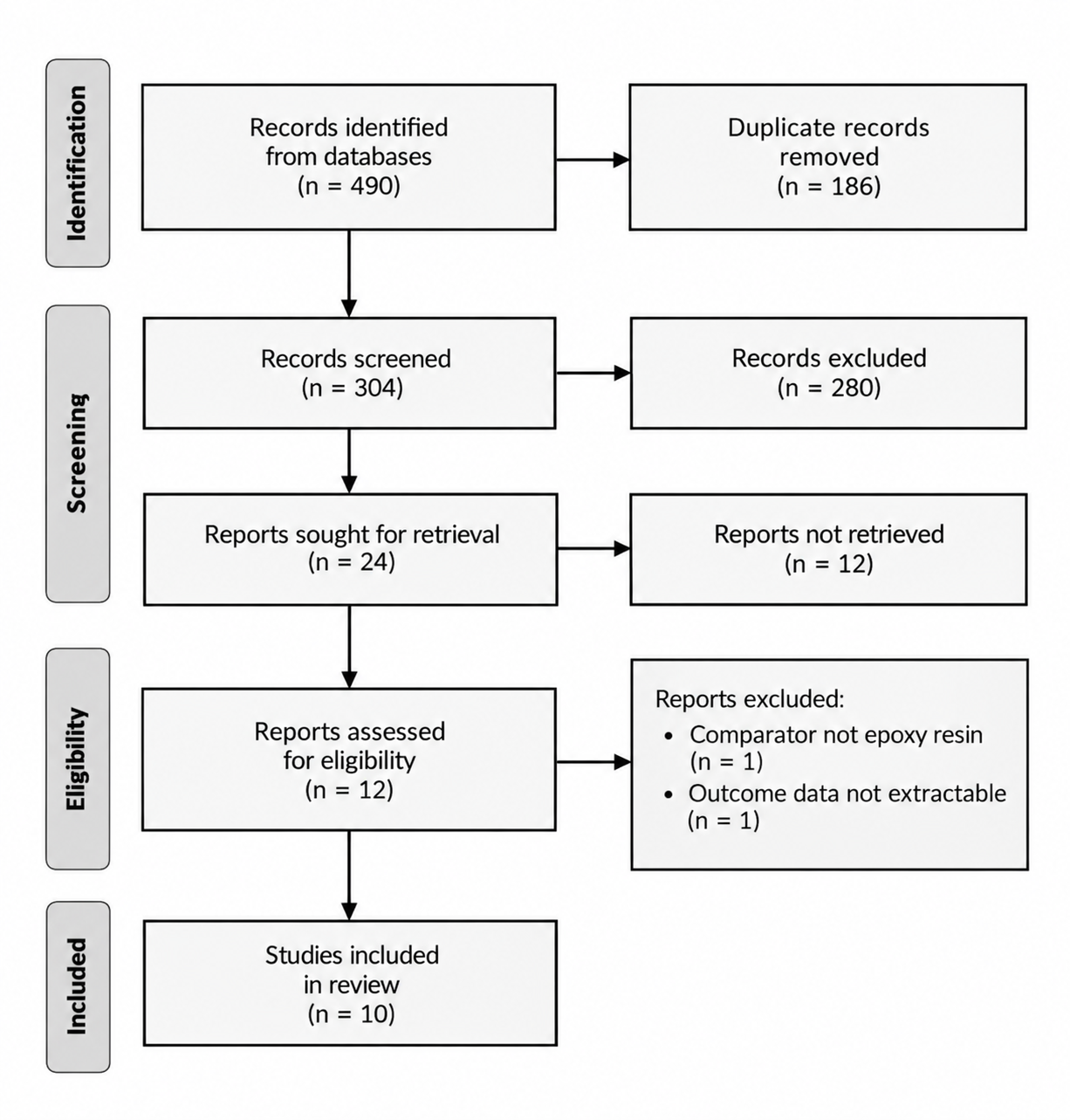
Figure 1. PRISMA 2020 flow diagram of study selection.
Note: The two reports excluded at full text were excluded because the comparator was not an epoxy resin sealer (n = 1) and because outcome data were not extractable (n = 1). Source: Elaborated from the search and screening records.Characteristics of included studies
The included studies comprised seven randomized trials,( 19-25) two prospective cohorts(26,27) and one non-randomized clinical trial,( 28) published between 2019 and 2023 in eight countries (Table 1). The bioceramic arm used EndoSequence BC Sealer, MTA Fillapex, BioRoot RCS, Endoseal MTA, Sealer Plus BC, Ceraseal or EndoSeal TCS; the epoxy arm used AH Plus in all studies, with ADseal as a second epoxy comparator in one trial.( 25) Five studies measured pain only, three measured healing only, and two measured both outcomes. Follow-up ranged from 5 days, for the pain trials, to 24 months for the healing cohorts. Sample sizes ranged from 60 to 150 teeth. The bioceramic sealers were generally applied with a single-cone or sealer-based technique, whereas AH Plus was applied with warm vertical compaction or continuous wave condensation, which couples the sealer comparison with the obturation technique in most studies. The treated teeth were predominantly molars and premolars, and the pulp and periapical diagnoses ranged from asymptomatic irreversible pulpitis to pulp necrosis with apical periodontitis; two studies included retreatment cases,(26,27) and one trial restricted inclusion to maxillary single-rooted teeth.( 24)
Study | Country | Design | Teeth (n) | Bioceramic sealer | Epoxy sealer | Outcomes | Follow-up |
|---|---|---|---|---|---|---|---|
Beshir 2021 | Egypt | RCT | 68 | EndoSequence BC | AH Plus | Pain (VAS) | 5 days |
Ferreira 2020 | Brazil | RCT | 60 | MTA Fillapex | AH Plus | Pain, analgesic | 7 days |
Khandelwal 2022 | India | RCT | 63 | BioRoot RCS | AH Plus | Pain, healing | 6 months |
Hussaen 2022 | Iraq | RCT | 75 | MTA Fillapex | AH Plus | Pain, analgesic | 7 days |
Shim 2021 | Korea | RCT | 108 | Endoseal MTA | AH Plus | Pain (VAS) | 7 days |
Fonseca 2019 | Brazil | RCT | 64 | Sealer Plus BC | AH Plus | Pain, extrusion | 1 week |
Song 2022 | Korea | RCT pilot | 80 | CeraSeal, EndoSeal TCS | AH Plus, ADseal | Pain, healing | 3 months |
Gautam 2022 | Nepal | Cohort | 100 | Bioceramic (SCO) | AH Plus (WVC) | Healing | 6-18 months |
Zamparini 2023 | Italy | Cohort | 94 | Ceraseal | AH Plus | Healing, survival | 24 months |
Zavattini 2020 | United Kingdom | NRCT | 150 | BioRoot RCS | AH Plus | Success (CBCT) | 12 months |
Note: RCT = randomized clinical trial; NRCT = non-randomized clinical trial; VAS = visual analog scale; CBCT = cone-beam computed tomography; SCO = single-cone obturation; WVC = warm vertical compaction. Source: Elaborated from the full texts of the included studies.
Description of the included studies
Beshir et al.(19) randomized 68 patients with asymptomatic apical periodontitis in multirooted teeth to EndoSequence BC Sealer or AH Plus with warm vertical compaction in a single visit, and recorded visual analogue scale pain from 6 hours to 5 days; pain was low and comparable between groups at every time point. Ferreira et al.(20) randomized 60 single-rooted teeth with pulp necrosis to AH Plus, Endofill or MTA Fillapex in a two-visit protocol, and found no difference in categorical pain or analgesic intake at 24 hours, 48 hours or 7 days. Khandelwal et al.(21) randomized 63 teeth with necrotic pulp and apical periodontitis to Tubli-Seal, AH Plus or BioRoot RCS, and reported the lowest 24-hour pain and the largest six-month reduction of the periapical lesion with BioRoot RCS. Hussaen et al.(22) randomized 75 single-rooted teeth to MTA Fillapex, CPM Sealer or AH Plus and found no difference in ordinal pain or analgesic intake. Shim et al.(23) randomized 108 patients, stratified by tooth type, to Endoseal MTA with single-cone obturation or AH Plus with continuous wave condensation, and found equivalent pain over 7 days but a shorter obturation time with the bioceramic sealer in molars. Fonseca et al.(24) randomized 64 maxillary single-rooted teeth to Sealer Plus BC or AH Plus with a single-cone technique, and reported similar pain despite more frequent extrusion of the bioceramic sealer. Song et al.(25) randomized 80 teeth to two epoxy sealers (AH Plus and ADseal) and two calcium silicate sealers (CeraSeal and EndoSeal TCS) in a sealer-based obturation, and observed negligible pain and comparable three-month healing in all groups. Gautam et al.(26) followed 100 teeth obturated with a bioceramic sealer and a single cone or with AH Plus and warm vertical compaction, and found more complete healing in the bioceramic group. Zamparini et al.(27) followed 94 teeth obturated with Ceraseal or AH Plus over Thermafil carriers and reported similar 24-month healing and survival. Zavattini et al.(28) treated 150 teeth with BioRoot RCS and a single cone or with AH Plus and warm vertical compaction, and reported similar 12-month success on cone-beam computed tomography.
Risk of bias
All seven randomized trials were judged at some concerns of bias, mainly because the operator could not be blinded to the sealer, which differed in presentation and handling; three trials additionally reported unclear or incomplete outcome data. None of the randomized trials was judged at low risk across all domains. Four trials( 19-21,24) used concealed randomization but could not blind the operator, and the remaining three(22,23,25) gave fewer details on allocation concealment or reported losses to follow-up. The two cohort studies and the non-randomized trial were judged at high risk of bias because allocation was not randomized: Gautam et al.( 26) and Zamparini et al.( 27) allocated by technique or operator training, and Zavattini et al.( 28) used an alternating sequence, so the groups could differ in baseline prognosis (Figure 2). No study was excluded on the basis of risk of bias, but the observational estimates were interpreted with this limitation in mind.

Figure 2. Risk-of-bias summary of the included studies.
Note: D1-D5 correspond to the domains of the Cochrane Risk of Bias 2 tool; observational studies were judged at high risk. Source: Elaborated from the included studies.Postoperative pain
Seven studies reported postoperative pain (Table 2). None found a statistically significant difference in pain incidence or intensity at any of the time points examined, from 6 hours to 7 days. The pain curves were similar in shape: in Beshir et al.,( 19) mean pain at 6 hours was 13.24 ± 23.83 in the EndoSequence BC group and 13.53 ± 24.48 in the AH Plus group, falling to 0.59 ± 2.39 and 5.29 ± 17.1, respectively, at 24 hours, with no significant difference at any point. Khandelwal et al.( 21) reported lower 24-hour scores with BioRoot RCS than with AH Plus (4.73 ± 7.72 vs 11.57 ± 11.18), converging to identical scores by 48 hours and to zero by 72 hours, and Fonseca et al.( 24) recorded pain of 1.21 ± 2.09 versus 1.46 ± 1.96 at 24 hours, falling to near zero by 48 hours in both groups. The same author recorded more frequent sealer extrusion with the bioceramic sealer (59.37% vs 28.12%) but no correlation between extrusion and pain. Ferreira et al.( 20) and Hussaen et al.( 22) measured pain as ordinal categories and found no difference in incidence, intensity or analgesic intake, and Shim et al.( 23) and Song et al.( 25) found near-zero pain scores regardless of the sealer. Analgesic consumption and flare-up rates were equally low across the two sealer families wherever they were reported. Three studies provided means and standard deviations at 24 hours and were pooled. The standardized mean difference was −0.36 (95% CI −0.66 to −0.05; p = 0.021; I² = 2.30%), indicating slightly lower 24-hour pain with bioceramic sealers, although the estimate was imprecise and rested on three small trials with low statistical power (Figure 3).
Study | Time point | Bioceramic mean ± SD (n) | Epoxy mean ± SD (n) | Finding |
|---|---|---|---|---|
Beshir 2021 | 24 h | 0.59 ± 2.39 (34) | 5.29 ± 17.1 (34) | No significant difference |
Fonseca 2019 | 24 h | 1.21 ± 2.09 (32) | 1.46 ± 1.96 (32) | No significant difference |
Khandelwal 2022 | 24 h | 4.73 ± 7.72 (21) | 11.57 ± 11.18 (21) | Lower with BioRoot RCS |
Ferreira 2020 | 24 h-7 d | Categories 1-4 | Categories 1-4 | No significant difference |
Hussaen 2022 | 24 h-7 d | Categories 1-4 | Categories 1-4 | No significant difference |
Shim 2021 | 7 d | VAS over 7 days | VAS over 7 days | No significant difference |
Song 2022 | 1 week | 0-0.06 | 0-0.06 | No significant difference |
Note: SD = standard deviation; VAS = visual analog scale; n = number of teeth or patients per group. Source: Elaborated from the full texts of the included studies.
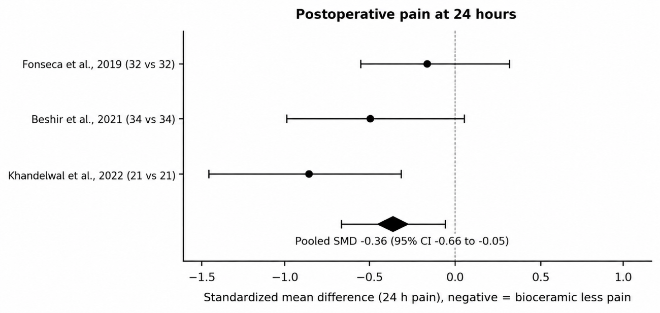
Figure 3. Forest plot of postoperative pain at 24 hours (standardized mean difference).
Note: Negative values favor the bioceramic sealer. Pooled estimate from a random-effects model. Source: Elaborated from the included studies.Periapical healing
Five studies reported radiographic healing (Table 3). Gautam et al.( 26) observed complete healing in 35 of 50 teeth (70%) obturated with a bioceramic sealer versus 24 of 50 (48%) obturated with AH Plus (p = 0.03), and the Kaplan-Meier analysis in that cohort showed a higher probability of healing in the bioceramic group as follow-up extended beyond 12 months. The remaining studies found similar success rates. Zavattini et al.( 28) reported 12-month success on cone-beam computed tomography in 45 of 53 (84%) bioceramic teeth versus 41 of 51 (80%) epoxy teeth (p = 0.6099), and 90% versus 89% on periapical radiographs (p = 0.7582). Zamparini et al.( 27) reported 24-month healing in 91.1% versus 88.6% (p = 0.624), with three of the six extruded Ceraseal masses becoming radiographically undetectable over follow-up. Khandelwal et al.( 21) described a larger mean reduction in lesion size with BioRoot RCS than with AH Plus at 6 months (10.22 vs 9.80), and Song et al.( 25) found similar radiographic regression of the periapical lesion at 3 months (33.3% vs 38.5%). The pooled risk ratio for the two studies that reported binary success was 1.04 (95% CI 0.93-1.16; p = 0.497; I² = 0.00%), indicating no difference in healing between the sealer families (Figure 4). Loss to follow-up was low in the healing studies, and tooth survival was high; Zamparini et al. reported a 24-month survival of 97.8% with one extraction in each group, both due to root fracture rather than endodontic failure.( 27)
Study | Follow-up | Bioceramic healing | Epoxy healing | Finding |
|---|---|---|---|---|
Gautam 2022 | 6-18 months | Complete healing 35/50 (70%) | Complete healing 24/50 (48%) | Higher with bioceramic (p = 0.03) |
Zavattini 2020 | 12 months | CBCT success 45/53 (84%) | CBCT success 41/51 (80%) | No significant difference (p = 0.6099) |
Zamparini 2023 | 24 months | Healed 41/45 (91.1%) | Healed 39/44 (88.6%) | No significant difference (p = 0.624) |
Khandelwal 2022 | 6 months | Lesion reduction 10.22 | Lesion reduction 9.80 | Greater with BioRoot RCS |
Song 2022 | 3 months | Regression 6/18 (33.3%) | Regression 5/13 (38.5%) | No significant difference |
Note: CBCT = cone-beam computed tomography; PAI = periapical index; n = number of teeth. Source: Elaborated from the full texts of the included studies.
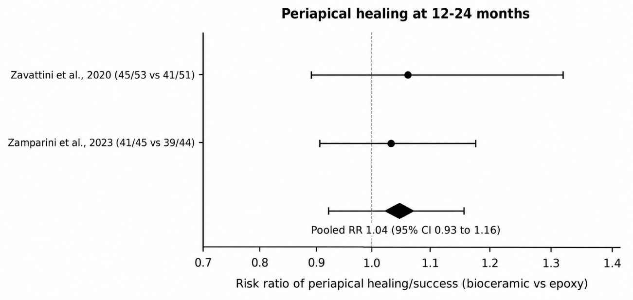
Figure 4. Forest plot of periapical healing at 12-24 months (risk ratio).
Note: Values above 1 favor the bioceramic sealer. Pooled estimate from a random-effects model. Source: Elaborated from the included studies.Secondary observations
Two secondary observations emerged from the included studies. Sealer extrusion was reported in 59.37% of the bioceramic group versus 28.12% of the epoxy group in one trial (Sealer Plus BC vs AH Plus),( 24) but in 13.3% of the Ceraseal group versus 25% of the AH Plus group in the cohort that used a warm carrier-based technique,( 27) and in neither case was extrusion associated with postoperative pain or with a worse healing outcome. Obturation time was shorter with the bioceramic sealer placed with a single cone than with AH Plus placed with continuous wave condensation in molars (198 vs 265 seconds).( 23) These observations reflect the handling characteristics of the materials rather than their biological activity, but they are relevant to the clinical choice between the two families.
Synthesis and certainty of the evidence
The pain meta-analysis was statistically consistent across the three trials (I² = 2.30%), but the confidence interval of the pooled standardized mean difference included small effects in both directions and the estimate was driven by two small trials; the certainty of the evidence for pain was therefore rated low, downgraded for imprecision and risk of bias. The three trials pooled for pain all used premixed, resin-free calcium silicate sealers (Sealer Plus BC, EndoSequence BC Sealer and BioRoot RCS), so the estimate does not include MTA-containing formulations, and the two MTA Fillapex trials could not be pooled because they reported pain as ordinal categories. The healing meta-analysis was also consistent (I² = 0.00%), but it rested on only two studies, its confidence interval crossed the null, and one of the two studies was observational; the certainty for healing was rated low for the same reasons. The two studies pooled for healing defined success as healed teeth, whereas Gautam et al. reported complete healing as a stricter endpoint and Khandelwal et al. reported lesion reduction as a continuous measure, which is why only two studies entered this analysis. Publication bias could not be assessed formally with so few studies, and the selective availability of open-access full texts is a further source of uncertainty that is quantified in the flow diagram. Taken together, the two analyses are consistent with clinical equivalence between the sealer families, with a possible small advantage for the bioceramic class limited to the first postoperative day.
DISCUSSION
In this systematic review, bioceramic and epoxy resin sealers produced broadly similar clinical outcomes. The pooled estimate pointed to slightly lower 24-hour postoperative pain with bioceramic sealers, but the difference was small, imprecise and driven by two of the three trials; periapical healing did not differ between the families at 12 to 24 months. The certainty of this evidence is low, and neither outcome provides a decisive argument for changing the sealer on clinical grounds alone; the direction of the pain estimate should be read as a signal that requires confirmation rather than as a demonstrated benefit.
The absence of a clear pain difference is consistent with individual trials that found no effect of the sealer on pain incidence or intensity,( 9,29-33) including the randomized comparisons pooled here,(23,24) and with studies that standardized instrumentation and excluded symptomatic teeth.( 3) The finding extends to retreatment: Ghobashy et al.( 33) found no difference in postoperative pain after single-visit retreatment with a bioceramic versus an epoxy resin sealer. Taken together, these comparisons indicate that when the technique and the clinical protocol are held constant, the sealer contributes little to the acute pain experience. This pattern is expected if the sealer is only one of several drivers of postoperative pain. Apical enlargement and pre-existing periapical disease influence postoperative pain more strongly than the sealer,(34,35) and when these factors are controlled, any material-specific effect may be too small to detect. The pain scores in the included trials were low and resolved within days, which also limits the room for a between-group difference to emerge.
Other syntheses reached a more favourable conclusion for bioceramic sealers, and the contrast requires explanation. Mekhdieva et al.( 11) reported significantly lower pain with bioceramic sealers at 24 and 48 hours, Seron et al.( 12) and Mendes et al.( 16) reported trends in the same direction, and Mahajan et al.( 18) described a modest level of evidence, whereas Sponchiado Junior et al.( 13) and Jamali et al.( 14) found no reduction in risk or intensity. The disagreement is explained by differences in the studies pooled rather than by any genuine inconsistency in the data. The reviews differ in the time point analysed, the pain scale used (visual analogue scales of 0 to 10 versus 0 to 100 versus ordinal categories), the sealer formulations grouped as bioceramic, and the inclusion of non-epoxy comparators. Ates et al.( 10) illustrate the point directly: pain scores were similar between iRoot SP and AH Plus, but analgesic intake was lower with the bioceramic sealer, so the choice of outcome determines the message. A review that pools analgesic intake will conclude in favour of the bioceramic sealer, while one that pools pain intensity will not.
For healing, the pooled risk ratio near 1 agrees with reviews that found comparable radiographic outcomes between the sealer families(15,17) and with observational data reporting high success rates for bioceramic sealers used with warm or single-cone techniques.(36,37) One retrospective comparison reported higher healing rates with a bioceramic sealer,(38) and the cohort of Gautam et al.(26) showed more complete healing with the bioceramic sealer; both were observational and confounded by the obturation technique, because the bioceramic arms used single-cone obturation while the epoxy arms used warm vertical compaction. Healing after root canal treatment is dominated by preoperative status, the presence of a lesion and the type of treatment rather than by the sealer,( 39-41) which probably explains why the sealer family contributes little to the final radiographic outcome once the canal is adequately cleaned and sealed. The consistency of the two pooled healing studies, which used different obturation carriers and follow-up periods and still produced risk ratios close to 1, is reassuring and suggests that the null finding is not an artefact of a single protocol.
The present meta-analyses update and extend the earlier ones. Sponchiado Junior et al.( 13) pooled five randomized trials published up to November 2020 and concluded that calcium silicate sealers do not reduce the risk or intensity of pain; our estimate, based on three trials reporting 24-hour means, is compatible with that null conclusion because the confidence interval nearly touches zero. Mekhdieva et al.( 11) and Seron et al.,( 12) by pooling a slightly different set of trials and time points, obtained a significant advantage for the bioceramic class; the discrepancy is a matter of which trials and which time point enter the analysis, not of different data. For healing, Chopra et al.( 15) and Khandelwal et al.( 17) likewise found no meaningful difference, and our pooled risk ratio of 1.04 with a confidence interval spanning 0.93 to 1.16 adds little to that conclusion.
Three sources of heterogeneity deserve attention because they could hide or exaggerate a sealer effect. The first is the grouping of the intervention. MTA Fillapex contains a salicylate resin in addition to mineral trioxide aggregate and behaves differently from premixed, resin-free calcium silicate sealers such as BioRoot RCS, Ceraseal or EndoSequence BC Sealer; pooling them as one bioceramic class may dilute or distort the estimate. The second is the coupling of the sealer with the obturation technique: in most included studies the bioceramic sealer was placed with a single cone or a sealer-based technique while AH Plus was placed with warm vertical compaction, so the comparison is between two complete obturation systems rather than between two sealers in isolation. The third is the definition and timing of the outcomes, which ranged from pain categories at 6 hours to cone-beam success at 24 months. These differences, more than a genuine biological effect, most plausibly explain the mixed results reported across the literature.(7,15) The MTA Fillapex question deserves particular attention because it sits at the boundary of the bioceramic definition: it combines mineral trioxide aggregate with a salicylate resin, is more soluble and less alkaline than premixed calcium silicate sealers, and is sometimes classified with resin sealers rather than with bioceramic ones.(4) Its two trials in this review reported categorical pain only, so its influence on the pooled pain estimate cannot be tested, but future analyses should separate it from the resin-free calcium silicate formulations.
The coupling of sealer and technique also has practical implications for interpreting the pooled estimates.
The coupling of sealer and technique also has practical implications for interpreting the pooled estimates. Single-cone obturation with a bioceramic sealer is faster and technically simpler than warm vertical compaction, as shown by the shorter obturation time recorded by Shim et al.,( 23) and sealer-based obturation is increasingly taught for its reproducibility.( 25) If the two systems produce equivalent pain and healing, the choice can legitimately be made on these technical grounds. The evidence assembled here supports equivalence rather than superiority, but it cannot separate the sealer effect from the technique effect, and a factorial trial that crosses sealer family with obturation technique would settle this question.
The borderline pain difference, if real, is biologically plausible. Calcium silicate sealers release calcium ions, raise pH and are less cytotoxic than epoxy resin sealers, which leach monomers that can activate trigeminal nociceptors and release calcitonin gene-related peptide.(5,7,8) Sealer extrusion was more frequent with some bioceramic formulations in the included studies(24) but was not associated with postoperative pain,(42) which is consistent with the resorption of extruded calcium silicate material observed over time.(27) The alkaline pH of the hydrated calcium silicate also contributes an antibacterial effect that the epoxy resin class does not share,(6) although its clinical relevance beyond the disinfection achieved by instrumentation and irrigation is uncertain. If the bioceramic advantage is real, it operates through reduced chemical irritation in the first 24 hours, before the sealer has fully set, and disappears once the acute inflammatory phase resolves.
The findings apply to permanent teeth treated under controlled conditions, mostly in university clinics, by experienced operators, and with single- or two-visit protocols; the pain estimates may not transfer to general practice, where symptomatic teeth, acute abscesses and less standardized instrumentation are more common.(2) The healing estimates, which come largely from cohorts with careful case selection, are likewise optimistic relative to routine practice.(39,40) The relative comparison between the two sealer families is unlikely to be affected by this, however, because the same clinical setting applied to both arms in each study.
The strengths of this review are its joint quantification of the two outcomes, its inclusion of both randomized and observational designs, and its restriction to a single bibliographic window with a fully reproducible search record. All analyses were computed deterministically from the extracted data and can be rerun, and every reference was verified against Crossref before citation.
Several limitations temper these conclusions. First, only 10 studies met the eligibility criteria, and 12 otherwise eligible reports could not be retrieved because only open-access full texts were used; the pooled analyses therefore rest on three and two studies, respectively, and are imprecise. A future update should obtain these reports to test whether the estimates change. Second, the included observational studies carry a high risk of bias from non-randomized allocation, and the randomized trials could not blind the operator to the sealer; a network meta-analysis that separates sealer formulations and obturation techniques would reduce this confounding. Third, pain was measured on different scales and at different time points, so only a minority of studies could be pooled; a core outcome set for endodontic postoperative pain would improve comparability across trials. Fifth, healing was assessed over short to medium follow-up, from 3 to 24 months, and with different imaging criteria, from periapical radiographs to cone-beam computed tomography; standardized periapical index or cone-beam-based outcome definitions, and longer follow-up, are needed.(43,44) Sixth, the bibliographic window ended in 2023, so trials published after that date could alter the pooled estimates. Seventh, the search was limited to English and Spanish, so studies published only in other languages could have been missed. Reporting bias could not be excluded either, because several included trials omitted standard deviations or reported only graphical pain data, which is why those studies could not enter the pooled analysis.
For practice, the choice between a bioceramic and an epoxy resin sealer should rest on handling, obturation technique and cost rather than on expected differences in pain or healing. The slight, imprecise reduction in 24-hour pain with bioceramic sealers is not sufficient, on its own, to justify a change of sealer, and the healing data offer no reason to prefer one family over the other. For research, the priorities are a registered protocol, a core outcome set for postoperative pain that fixes the scale and the time points, radiographic healing assessed with a standardized periapical index or cone-beam computed tomography over at least 12 months, and trials that cross the sealer family with the obturation technique so that the two effects can be separated. Such trials would resolve the residual uncertainty with far less ambiguity than further small, heterogeneous comparisons.
CONCLUSIONS
Bioceramic and epoxy resin sealers produced similar postoperative pain after root canal treatment, with a small, imprecise reduction in 24-hour pain favouring bioceramic sealers in a pooled analysis of three trials. Periapical healing did not differ between the sealer families at 12 to 24 months. The certainty of the evidence is low, and further randomized trials with standardized pain and healing measures are needed before one sealer family can be recommended over the other.
References
- 1 Sathorn C, Parashos P, Messer H. The prevalence of postoperative pain and flare‐up in single‐ and multiple‐visit endodontic treatment: a systematic review. Int Endodontic J. 2008;41(2):91-99. doi: 10.1111/j.1365-2591.2007.01316.x
- 2 Gomes MS, Böttcher DE, Scarparo RK, Morgental RD, Waltrick SBG, Ghisi AC, et al. Predicting pre‐ and postoperative pain of endodontic origin in a southern Brazilian subpopulation: an electronic database study. Int Endodontic J. 2017;50(8):729-739. doi: 10.1111/iej.12684
- 3 Keskin C, Sivas Yilmaz Ö, Inan U, Özdemir Ö. Postoperative pain after glide path preparation using manual, reciprocating and continuous rotary instruments: a randomized clinical trial. Int Endodontic J. 2019;52(5):579-587. doi: 10.1111/iej.13053
- 4 KOMABAYASHI T, COLMENAR D, CVACH N, BHAT A, PRIMUS C, IMAI Y. Comprehensive review of current endodontic sealers. Dent Mater J. 2020;39(5):703-720. doi: 10.4012/dmj.2019-288
- 5 TROIANO G, PERRONE D, DIOGUARDI M, BUONAVOGLIA A, ARDITO F, LO MUZIO L. <i>In vitro</i> evaluation of the cytotoxic activity of three epoxy resin-based endodontic sealers. Dental Materials Journal. 2018;37(3):374-378. doi: 10.4012/dmj.2017-148
- 6 AL-Haddad A, Che Ab Aziz ZA. Bioceramic-Based Root Canal Sealers: A Review. International Journal of Biomaterials. 2016;2016:1-10. doi: 10.1155/2016/9753210
- 7 Camilleri J, Atmeh A, Li X, Meschi N. Present status and future directions: Hydraulic materials for endodontic use. Int Endodontic J. 2022;55(S3):710-777. doi: 10.1111/iej.13709
- 8 Wuersching SN, Diegritz C, Hickel R, Huth KC, Kollmuss M. A comprehensive in vitro comparison of the biological and physicochemical properties of bioactive root canal sealers. Clin Oral Invest. 2022;26(10):6209-6222. doi: 10.1007/s00784-022-04570-2
- 9 Graunaite I, Skucaite N, Lodiene G, Agentiene I, Machiulskiene V. Effect of Resin-based and Bioceramic Root Canal Sealers on Postoperative Pain: A Split-mouth Randomized Controlled Trial. Journal of Endodontics. 2018;44(5):689-693. doi: 10.1016/j.joen.2018.02.010
- 10 Atav Ates A, Dumani A, Yoldas O, Unal I. Post-obturation pain following the use of carrier-based system with AH Plus or iRoot SP sealers: a randomized controlled clinical trial. Clin Oral Invest. 2019;23(7):3053-3061. doi: 10.1007/s00784-018-2721-6
- 11 Mekhdieva E, Del Fabbro M, Alovisi M, Comba A, Scotti N, Tumedei M, et al. Postoperative Pain following Root Canal Filling with Bioceramic vs. Traditional Filling Techniques: A Systematic Review and Meta-Analysis of Randomized Controlled Trials. JCM. 2021;10(19):4509. doi: 10.3390/jcm10194509
- 12 Seron MA, Nunes GP, Ferrisse TM, Strazzi-Sahyon HB, Victorino FR, dos Santos PH, et al. Postoperative pain after root canal filling with bioceramic sealers: a systematic review and meta-analysis of randomized clinical trials. Odontology. 2023;111(4):793-812. doi: 10.1007/s10266-023-00830-z
- 13 Sponchiado Junior EC, Vieira WDA, Normando AGC, Pereira JV, Ferraz CCR, Almeida JFA, et al. Calcium Silicate-Based Sealers Do Not Reduce the Risk and Intensity of Postoperative Pain after Root Canal Treatment when Compared with Epoxy Resin-Based Sealers: A Systematic Review and Meta-Analysis. Eur J Dent. 2021;15(02):347-359. doi: 10.1055/s-0041-1724157
- 14 Jamali S, Darvish M, Nasrabadi N, Jafarizadeh S. Evaluation of the Effect of the Intensity and Occurrence of Postoperative Pain of Resin-Based and Bioceramic Root Canal Sealers: A Systematic Review and Meta-Analysis of Randomized Controlled Trial Studies. Pesqui Bras Odontopediatria Clín Integr. 2021;21:e0219. doi: 10.1590/pboci.2021.082
- 15 Chopra V, Davis G, Baysan A. Clinical and Radiographic Outcome of Non-Surgical Endodontic Treatment Using Calcium Silicate-Based Versus Resin-Based Sealers—A Systematic Review and Meta-Analysis of Clinical Studies. JFB. 2022;13(2):38. doi: 10.3390/jfb13020038
- 16 Teixeira Mendes A, Weissheimer T, Tietz L, Barcelos Só B, Barcelos Só G, Abreu da Rosa R, et al. Postoperative pain incidence and intensity following root canal obturation with bioceramic and other sealers: A systematic review with meta-analysis. J Res Dent. 2023;11(2):32-51. doi: 10.59306/jrd.v11e2202332-51
- 17 Khandelwal A, Janani K, Teja K, Jose J, Battineni G, Riccitiello F, et al. Periapical Healing following Root Canal Treatment Using Different Endodontic Sealers: A Systematic Review. BioMed Research International. 2022;2022(1):3569281. doi: 10.1155/2022/3569281
- 18 Mahajan D, Manocha D, Patel P, Saraiya MB, Chaniyara K. A Systematic Review on Comparison of Periapical Healing and Post-Operative Pain between Bioceramic and Epoxy Resin Based Sealers. J Pharm Bioallied Sci. 2023;15(Suppl 2):S862-S866. doi: 10.4103/jpbs.jpbs_145_23
- 19 Beshir MA, Sadek HS, El-Asfouri H. Single Visit Root Canal Treatment: Intensity of Postoperative Pain Using AH Plus Sealer Versus Endosequence BC Sealer in Patients with Asymptomatic Apical Periodontitis (A Randomized Clinical Trial). Act Scie Dental. 2021:102-113. doi: 10.31080/asds.2021.05.1264
- 20 FERREIRA NDS, GOLLO EKF, BOSCATO N, ARIAS A, SILVA EJNLD. Postoperative pain after root canal filling with different endodontic sealers: a randomized clinical trial. Braz oral res. 2020;34:e069. doi: 10.1590/1807-3107bor-2020.vol34.0069
- 21 Akshay K, Jerry J, Kavalipurapu-Venkata T, Ajitha P. Comparative evaluation of postoperative pain and periapical healing after root canal treatment using three different base endodontic sealers – A randomized control clinical trial. J Clin Exp Dent. 2022:e144-e152. doi: 10.4317/jced.59034
- 22 Hussaen MN, Abbas SMJ, Rahoomi MH. Evaluation of the effect of three different base endodontic sealers on the periapical region's discomfort and healing after root canal therapy. ijhs. 2022:2025-2034. doi: 10.53730/ijhs.v6ns8.12384
- 23 Shim K, Jang YE, Kim Y. Comparison of the Effects of a Bioceramic and Conventional Resin-Based Sealers on Postoperative Pain after Nonsurgical Root Canal Treatment: A Randomized Controlled Clinical Study. Materials. 2021;14(10):2661. doi: 10.3390/ma14102661
- 24 Fonseca B, Coelho MS, Bueno CEDS, Fontana CE, Martin ASD, Rocha DGP. Assessment of Extrusion and Postoperative Pain of a Bioceramic and Resin-Based Root Canal Sealer. Eur J Dent. 2019;13(03):343-348. doi: 10.1055/s-0039-3399457
- 25 Song M, Park MG, Kwak SW, Kim RH, Ha JH, Kim HC. Pilot Evaluation of Sealer-Based Root Canal Obturation Using Epoxy-Resin-Based and Calcium-Silicate-Based Sealers: A Randomized Clinical Trial. Materials. 2022;15(15):5146. doi: 10.3390/ma15155146
- 26 Gautam S, Thapa A, Joshi R, Joshi B. Outcome of root canal treatment using Bioceramic sealer and resinbased sealer: An observational analytical study. Nep Med Coll J. 2022;24(1):68-74. doi: 10.3126/nmcj.v24i1.44144
- 27 Zamparini F, Spinelli A, Cardinali F, Ausiello P, Gandolfi MG, Prati C. The Use of Premixed Calcium Silicate Bioceramic Sealer with Warm Carrier-Based Technique: A 2-Year Study for Patients Treated in a Master Program. JFB. 2023;14(3):164. doi: 10.3390/jfb14030164
- 28 Zavattini A, Knight A, Foschi F, Mannocci F. Outcome of Root Canal Treatments Using a New Calcium Silicate Root Canal Sealer: A Non-Randomized Clinical Trial. JCM. 2020;9(3):782. doi: 10.3390/jcm9030782
- 29 Aslan T, Dönmez Özkan H. The effect of two calcium silicate‐based and one epoxy resin‐based root canal sealer on postoperative pain: a randomized controlled trial. Int Endodontic J. 2021;54(2):190-197. doi: 10.1111/iej.13411
- 30 Yu YH, Kushnir L, Kohli M, Karabucak B. Comparing the incidence of postoperative pain after root canal filling with warm vertical obturation with resin-based sealer and sealer-based obturation with calcium silicate-based sealer: a prospective clinical trial. Clin Oral Invest. 2021;25(8):5033-5042. doi: 10.1007/s00784-021-03814-x
- 31 Coşar M, Kandemir Demirci G, Çalışkan MK. The effect of two different root canal sealers on treatment outcome and post‐obturation pain in single‐visit root canal treatment: A prospective randomized clinical trial. Int Endodontic J. 2023;56(3):318-330. doi: 10.1111/iej.13870
- 32 Kim JH, Cho SY, Choi Y, Kim DH, Shin SJ, Jung IY. Clinical Efficacy of Sealer-based Obturation Using Calcium Silicate Sealers: A Randomized Clinical Trial. Journal of Endodontics. 2022;48(2):144-151. doi: 10.1016/j.joen.2021.11.011
- 33 Ghobashy A, Fakhr M. Postoperative Pain evaluation after single visit nonsurgical retreatment using a Bio ceramic sealer. Egyptian Dental Journal. 2022;68(4):4029-4037. doi: 10.21608/edj.2022.155094.2207
- 34 de Freitas Portela FSM, De Martin AS, Pelegrine RA, Gutmann JL, Kato AS, Bueno CEDS. Effect of Foraminal Enlargement on Postoperative Pain in Necrotic Single-Rooted Teeth: A Randomized Clinical Trial. Journal of Endodontics. 2021;47(7):1046-1051. doi: 10.1016/j.joen.2021.04.008
- 35 AbdurRahman S, Abdel Aziz SM, Gawdat SI, AbdalSamad AM. Postoperative pain of patients with necrotic teeth with apical periodontitis following single visit endodontic treatment versus multiple visit endodontic treatment using triple antibiotic paste: a randomized clinical trial. F1000Res. 2019;8:1203. doi: 10.12688/f1000research.19936.1
- 36 Pontoriero DIK, Ferrari Cagidiaco E, Maccagnola V, Manfredini D, Ferrari M. Outcomes of Endodontic-Treated Teeth Obturated with Bioceramic Sealers in Combination with Warm Gutta-Percha Obturation Techniques: A Prospective Clinical Study. JCM. 2023;12(8):2867. doi: 10.3390/jcm12082867
- 37 Li J, Chen L, Zeng C, Liu Y, Gong Q, Jiang H. Clinical outcome of bioceramic sealer iRoot SP extrusion in root canal treatment: a retrospective analysis. Head Face Med. 2022;18(1):28. doi: 10.1186/s13005-022-00332-3
- 38 Alharith DN, Mansi IT, Abdulmotalib Y, Amous H, Aljulban T, Aiban HMA, et al. Radiographic Evaluation of Periapical Healing Rates Between Bio-Ceramic Sealer and AH+ Sealer: A Retrospective Study. Ann Dent Spec. 2023;11(2):124-128. doi: 10.51847/rtpmlou0gt
- 39 Eyuboglu TF, Olcay K, Özcan M. A clinical study on single-visit root canal retreatments on consecutive 173 patients: frequency of periapical complications and clinical success rate. Clin Oral Invest. 2017;21(5):1761-1768. doi: 10.1007/s00784-016-1957-2
- 40 Laukkanen E, Vehkalahti MM, Kotiranta AK. Impact of systemic diseases and tooth‐based factors on outcome of root canal treatment. Int Endodontic J. 2019;52(10):1417-1426. doi: 10.1111/iej.13143
- 41 Johnsen I, Bårdsen A, Haug SR. Impact of Case Difficulty, Endodontic Mishaps, and Instrumentation Method on Endodontic Treatment Outcome and Quality of Life: A Four-Year Follow-up Study. Journal of Endodontics. 2023;49(4):382-389. doi: 10.1016/j.joen.2023.01.005
- 42 Drumond JPSC, Maeda W, Nascimento WM, Campos DDL, Prado MC, de-Jesus-Soares A, et al. Comparison of Postobturation Pain Experience after Apical Extrusion of Calcium Silicate– and Resin–Based Root Canal Sealers. Journal of Endodontics. 2021;47(8):1278-1284. doi: 10.1016/j.joen.2021.05.008
- 43 Ørstavik D, Kerekes K, Eriksen HM. The periapical index: A scoring system for radiographic assessment of apical periodontitis. Dental Traumatology. 1986;2(1):20-34. doi: 10.1111/j.1600-9657.1986.tb00119.x
- 44 Zanini M, Decerle N, Hennequin M, Cousson P. Revisiting Orstavik’s PAI score to produce a reliable and reproducible assessment of the outcomes of endodontic treatments in routine practice. Eur J Dental Education. 2021;25(2):291-298. doi: 10.1111/eje.12603
Declarations
Funding
No financing
Conflict of interest
None
Authorship contributions
Drafting – original draft: Ernesto Learsys Campello Trujillo.
Writing–review and editing: Ernesto Learsys Campello Trujillo.