

Abstract
Introduction: Demarcated opacities of molar-incisor hypomineralization are a frequent aesthetic concern in children and adolescents. Resin infiltration has been proposed as a minimally invasive alternative to microabrasion, bleaching and restorative approaches, but its comparative effectiveness remains unclear. Methods: A systematic review following PRISMA 2020 was conducted. PubMed, OpenAlex and Crossref were searched for clinical and image-based studies published between 2015 and 2023 that compared resin infiltration with other minimally invasive approaches for masking demarcated opacities in molar-incisor hypomineralization. Risk of bias was assessed with RoB 2 and ROBINS-I, and findings were synthesized narratively. Results: Of 237 records, 10 studies were included. Resin infiltration reduced the color difference between the opacity and sound enamel in every comparative study; the difference was 4.07 units after infiltration versus 7.35 after placebo, and resin infiltration outperformed fluoride varnish at all time points. Complete masking was rare, and partial masking predominated, reported in 95.65% of infiltrated teeth in one study. Most studies were small, short and at serious risk of bias. Conclusions: Resin infiltration reduces but rarely fully conceals demarcated opacities in molar-incisor hypomineralization, with short-term results generally superior to placebo and fluoride varnish. Direct randomized comparisons against microabrasion are lacking, and the evidence does not yet support one protocol over another.
Keywords
molar incisor hypomineralization, dental enamel hypomineralization, resin infiltration, dental enamel, esthetics dental, minimally invasive dentistry
INTRODUCTION
Molar-incisor hypomineralization (MIH) is a qualitative enamel defect of systemic origin that affects one to four first permanent molars and is frequently accompanied by involvement of the permanent incisors.(1) Its pooled prevalence has been estimated around 14% in the general child population, with substantial variation across regions and diagnostic criteria.(2,3) Diagnosis is based on the European Academy of Paediatric Dentistry criteria, which define the condition by the presence of demarcated opacities, posteruptive enamel breakdown, atypical restorations or the extraction of affected first molars, and a standardized scoring method has been proposed to make epidemiological records comparable.(4) On the labial surfaces of anterior teeth the defect appears as well-demarcated opacities whose color ranges from creamy white to yellow-brown, and these opacities are among the most common reasons why children and their parents seek aesthetic treatment.(5,6) The clinical burden extends beyond appearance, because affected teeth are also hypersensitive, prone to rapid caries progression and difficult to restore, which together make MIH one of the more demanding conditions in paediatric practice.(7,8) The visual impact is likewise not merely cosmetic: visible opacities have been associated with reduced oral health-related quality of life and with negative self-perception during a developmental period in which self-image is being consolidated.(5,6)
The condition is graded by the extension and color of the lesions and by the presence of posteruptive breakdown, and the treatment decision follows that severity: mild opacities can be monitored, whereas visible incisor opacities and broken molars require progressively more invasive management.(9) For anterior opacities in particular the treatment aim shifts from protection to appearance, and the clinician must balance the benefit of concealment against the amount of enamel that each technique sacrifices.(10) This tension, between a predictable appearance and the preservation of tooth structure, is the practical context in which resin infiltration is proposed.
Clinical management of MIH opacities spans a wide spectrum. Mild lesions can be observed or treated with remineralizing agents, whereas more visible defects are managed with enamel microabrasion, tooth bleaching, resin infiltration or direct composite veneers, and severe lesions may require full-coverage restorations.(9,10) Resin infiltration occupies an intermediate position because it removes only a thin hypermineralized surface layer with hydrochloric acid, replaces water in the porous lesion body with ethanol and then fills the intercrystalline spaces with a low-viscosity resin whose refractive index approaches that of sound enamel.(11,12) The marketed Icon system standardizes this sequence with a 15% hydrochloric acid gel applied for two minutes, ethanol dehydration for thirty seconds and two consecutive applications of the infiltrant, each followed by light polymerization.(13) The procedure is completed in a single visit, requires no anaesthesia in most cases and preserves enamel tissue, which makes it attractive for children.(10) Its optical effect, however, depends on the depth and the organic content of the lesion, and MIH enamel is protein-rich and more resistant to penetration than the demineralized enamel of white spot lesions, so the aesthetic response may be less predictable.(11) In children, the choice among these options is further shaped by behavior management, by the wish to defer irreversible restorative procedures while the enamel is still maturing, and by the principle that the least destructive treatment should be attempted before more invasive ones, all of which favor microabrasion, bleaching and infiltration over direct veneering in mild to moderate defects.(9,10)
Previous syntheses have examined resin infiltration either in enamel defects in general or within broader overviews of MIH treatment.(14-17) Reviews restricted to resin infiltration in MIH concluded that the technique improves the appearance of demarcated opacities, but they relied on small uncontrolled series and did not isolate the comparative question of how infiltration performs against other minimally invasive options.(14,18) An overview of systematic reviews on MIH treatment judged the overall quality of the available evidence as low, and a meta-analysis restricted to white spot lesions and fluorosis cannot be transferred directly to MIH because the two substrates differ histologically.(19,20) The reviews closest to the present topic assembled mostly case reports and short series, and the broader treatment reviews were organized by clinical presentation rather than by the comparative aesthetic question, so none of them quantified how infiltration performs against the alternative minimally invasive options.(14,15,17) It therefore remains unclear whether resin infiltration should be preferred over microabrasion, bleaching or fluoride varnish when the clinical objective is to conceal a demarcated MIH opacity.
Although the optical principle of infiltration is the same regardless of etiology, the response of MIH opacities is not interchangeable with that of caries-related white spot lesions or fluorosis. MIH enamel retains a higher protein content and a porous structure that differs in depth and distribution from demineralized enamel, and yellow-brown opacities in particular tend to be deeper and less permeable to the infiltrant.(11) Evidence from fluorosis and white spot lesions therefore cannot be extrapolated directly to MIH, and the question of whether infiltration conceals MIH opacities as effectively as it conceals other enamel defects, or as effectively as microabrasion does, has to be answered with studies conducted in the MIH population itself.(20)
The aim of this systematic review was to compare the aesthetic effectiveness of resin infiltration versus other minimally invasive approaches for masking demarcated enamel opacities in patients with molar-incisor hypomineralization. The review addressed the question: in patients with molar-incisor hypomineralization, how effective is resin infiltration in masking demarcated opacities compared with other minimally invasive approaches?
METHODS
Protocol and registration
This systematic review was planned and reported according to the PRISMA 2020 statement.(21) A protocol was not prospectively registered in PROSPERO or any other registry, which is reported here as a deviation from current best practice.
Eligibility criteria
Studies were selected according to a PICO framework. The population comprised children and adolescents with a clinical diagnosis of molar-incisor hypomineralization and demarcated opacities on permanent anterior teeth; studies restricted to fluorosis, caries-associated white spot lesions or other developmental defects were excluded unless they reported MIH separately. The intervention was resin infiltration, applied with the Icon system or an equivalent low-viscosity infiltrant, following the standard sequence of hydrochloric acid etching, ethanol dehydration and resin application. The comparator was any other minimally invasive approach intended to improve the appearance of the opacity, including enamel microabrasion, tooth bleaching, fluoride varnish, remineralizing agents and placebo or no treatment. The outcome was the masking effect, expressed as the color difference between the opacity and adjacent sound enamel, the visible opacity area or brightness, or a categorical assessment of concealment; studies reporting only hypersensitivity, structural integrity or surface roughness were not eligible. Randomized controlled trials, non-randomized comparative studies, uncontrolled before-after clinical studies and image-based analyses of treated patients were eligible; case reports, laboratory studies on extracted teeth and reviews were excluded. Only records published between 2015 and 2023 were considered, and no language restriction was applied at the search stage, provided that an English abstract was available for screening.
Information sources and search
PubMed, OpenAlex and Crossref were restricted to the 2015 to 2023 publication window. The following string illustrates the strategy applied in OpenAlex: ("molar incisor hypomineralization" OR "molar-incisor hypomineralisation" OR "enamel hypomineralization") AND ("resin infiltration" OR "infiltration" OR "microabrasion" OR "micro-abrasion" OR "minimally invasive") AND ("masking" OR "opacity" OR "opacities" OR "aesthetic" OR "esthetic" OR "color"). Variants of this string were adapted to each database and combined with terms for white spot lesions and developmental enamel defects to capture comparator studies. In addition, a targeted search for comparative masking studies was run, and reference lists of the included reviews were screened for primary studies. A core set of searches populated the records screened for eligibility, and a second set of supporting searches, run with the same window but without the MIH restriction, retrieved the literature used for context and for the discussion of mechanism and comparators. The supporting searches were intended only to locate comparator and mechanism literature and did not alter the set of included studies, which was defined exclusively by the core search results and their screening.
Selection process
Records were deduplicated and screened in two stages. In the first stage, titles and abstracts were assessed against the eligibility criteria. Records judged potentially relevant or uncertain proceeded to the second stage, in which eligibility was confirmed using the available full text or, when the full text was not openly accessible, the structured abstract retrieved from the database. Screening and data extraction were assisted by an artificial intelligence system and verified by the author; a single reviewer performed the selection rather than two independent reviewers, and this is acknowledged as a source of potential bias. Disagreements between the reviewer and the automated suggestion were resolved by the reviewer. Exclusion reasons were coded at the full-text stage using predefined categories covering population, intervention, outcome, design and record verifiability, and the counts of each reason were carried forward into the flow diagram. No hand searching of grey literature was performed and conference abstracts were not sought, so the review may miss unpublished comparative studies.
Data extraction
For each included study the following were extracted into a structured form: first author and year, country, design, sample size, age range, intervention and comparator, outcome measure, the main result with its estimate, and follow-up. Records were managed in structured files that preserved the source of each value, and the selection counts were derived from those records rather than transcribed by hand, so that the flow diagram and the reference list remained consistent. Each datum was recorded with its source location. When only the abstract was available, extraction was limited to the information reported there and fields were not completed by inference. Where the full text was openly accessible, quantitative values were transcribed directly from the results section, including means, standard deviations and the reported statistical tests, and no value was recalculated or imputed. Color outcomes were retained in the units reported by each study, most commonly the CIELAB color difference, the change in lightness, a greyscale brightness value, or a categorical classification of the degree of concealment.
Risk of bias
Randomized trials were assessed with the Cochrane risk-of-bias tool for randomized trials (RoB 2),(22) and non-randomized studies with the Risk Of Bias In Non-randomized Studies of Interventions (ROBINS-I) tool.(23) For randomized trials the domains considered were the randomization process, deviations from intended interventions, missing outcome data, measurement of the outcome and selection of the reported result. For non-randomized studies the domains were confounding, selection of participants, classification of interventions, deviations from intended interventions, missing data, measurement of outcomes and selection of the reported result. Because most records could be assessed only from their abstracts, domains that could not be verified from the available text were rated as some concerns or no information rather than low risk. The image-based study was not a trial and was evaluated qualitatively for measurement reliability rather than with a trial risk-of-bias tool, and its intraclass correlation coefficients were recorded as an indicator of measurement repeatability.
Synthesis
Findings were synthesized narratively and grouped by the type of comparison: resin infiltration without a concurrent comparator, resin infiltration versus control, placebo or fluoride varnish, and resin infiltration or microabrasion used alone or in combination. A meta-analysis was not performed because the studies differed in design, comparator, outcome metric and follow-up, and only one or two studies were available for any single comparison; pooling such heterogeneous estimates was considered inappropriate. Effect estimates are therefore reported as they appeared in each study rather than as a single summary measure. A formal assessment of the certainty of the evidence, such as the GRADE approach, was not undertaken because the evidence base did not allow a coherent outcome-by-outcome grading; the overall low confidence in the findings is instead described qualitatively. The possibility of reporting bias could not be examined with funnel plots given the small number of studies per comparison, and this limitation is acknowledged in the interpretation.
Deviations and transparency
Several aspects of the execution differed from the ideal systematic review and are reported here rather than concealed. Full-text retrieval was limited to openly accessible records, so eligibility and data extraction for the remaining studies relied on the structured abstracts retrieved from the databases, which prevented a complete assessment of the risk-of-bias domains and restricted extraction to the values that authors reported in the abstract. Screening was performed by a single reviewer with automated assistance rather than by two independent reviewers, and disagreements were resolved by the reviewer. The protocol was not prospectively registered, and the certainty of the evidence was described qualitatively instead of being graded with the GRADE framework. These choices are declared as limitations and should be taken into account when the review is interpreted.
RESULTS
Study selection
The searches identified 237 records, of which 47 were duplicates, leaving 190 records for title and abstract screening (Figure 1). Of these, 176 were excluded. The exclusions at this stage consisted mainly of narrative and systematic reviews, prevalence and etiology surveys, case reports and case series, laboratory studies on extracted teeth, and studies whose population was fluorosis, caries-associated white spot lesions or post-orthodontic demineralization rather than molar-incisor hypomineralization. The remaining 14 reports were assessed for eligibility, and 4 were excluded: one because the outcome was structural integrity rather than masking, one because the outcome was surface roughness and hypersensitivity, and two because their bibliographic records could not be verified against a resolvable identifier. Finally, 10 studies were included in the review.
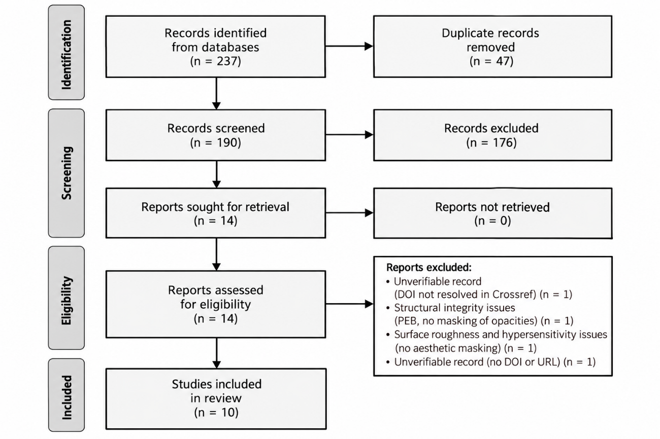
Figure 1. PRISMA 2020 flow diagram of study selection.
Note: The diagram reports the records identified from the databases, those excluded at each stage and the studies finally included. Source: Elaborated from the screening records of this review.Characteristics of the included studies
The 10 included studies were published between 2017 and 2023 and are summarized in Table 1 (Table 1). Three were randomized controlled trials, one was a non-randomized controlled study, one was a non-randomized comparative study, and the remaining five were uncontrolled before-after clinical studies, one of which analyzed digital clinical photographs of treated patients. Sample sizes ranged from 20 to 39 patients and from 22 to 116 teeth, and participants were aged between 7 and 18 years; where sex was reported, the samples included both girls and boys.(13) The studies were conducted in India, Turkey, Italy, Poland, Egypt and Brazil. Resin infiltration was applied with the Icon system in all studies that named the product, and the comparators were placebo, fluoride varnish, microabrasion alone, and microabrasion combined with remineralizing agents. Follow-up ranged from one month to 24 months, with most studies reporting six months or less. Two records enrolled populations that were not restricted to MIH: Brescia et al. included mild and moderate fluorosis and post-traumatic hypomineralization alongside MIH, and Gençer and Kirzioğlu analyzed fluorosis and hypomineralization subgroups separately, so only the MIH-relevant findings from these records were considered here.(24,25) All infiltration procedures followed the sequence of hydrochloric acid etching, ethanol dehydration and resin application described by the manufacturer, and the outcome was measured with a spectrophotometer, with digital image analysis, or with a categorical clinical classification depending on the study.
Study (year) | Country | Design | Sample | Intervention vs comparison | Outcome | Follow-up |
|---|---|---|---|---|---|---|
Bhandari et al. (2018) | India | Before-after | 22 incisors | RI (Icon-type) | ΔE and ΔL | 6 months |
Elbaz and Omer (2017) | Egypt | RCT | 20 children | RI vs FV | ΔE and grey level | 1 month |
Bhandari et al. (2019) | India | RCT | 46 lesions | MA vs MA + CPP-ACFP | ΔL and ΔE | 6 months |
Gençer and Kirzioğlu (2019) | Turkey | Comparative | incisors, n not reported | RI vs MA + remineralizer | L* and ΔE | not reported |
Brescia et al. (2022) | Italy | Retrospective | 33 patients | RI (Icon) | Aesthetic improvement | 24 months |
Athayde et al. (2022) | Brazil | RCT | 39 patients | RI vs placebo | ΔE and total masking | 1 month |
Warner et al. (2022) | United Kingdom | Image-based | 23 patients | MA and/or RI | Opacity area and brightness | 6 months |
Ozgur et al. (2023) | Turkey | Prospective | 84 teeth | RI | ΔE | 6 months |
Ćwiklińska et al. (2023) | Poland | Before-after | 115 incisors | RI (Icon) | Masking category | 6 months |
Altan and Yilmaz (2023) | Turkey | Non-randomized controlled | 116 incisors | RI vs no treatment (healthy) | ΔE, lesion area, DIAGNOdent | 6 months |
Note: RI = resin infiltration; MA = microabrasion; FV = fluoride varnish; RCT = randomized controlled trial; ΔE = color difference; n = sample size. Source: Elaborated from the data extracted from each included study.
Risk of bias within studies
Risk of bias is summarized in Table 2 (Table 2). The three randomized trials were rated as raising some concerns, mainly because the abstracts did not report how the allocation sequence was generated and concealed, and no full text was available to complete those domains. The non-randomized and uncontrolled studies were rated as serious or critical risk of bias, principally because they lacked a concurrent comparator group, so any change in appearance cannot be separated from natural variation, dehydration of the enamel during the procedure or examiner expectation. The controlled non-randomized study compared infiltrated MIH teeth with untreated healthy teeth, which is a source of confounding because the two groups differed in their baseline optical properties. Among the randomized trials, none of the abstracts reported the method of sequence generation or allocation concealment, and only Athayde et al. described a placebo control that would allow blinding of the participants and, to some extent, of the outcome assessor.(26) The uncontrolled series could not exclude regression to the mean or examiner expectation, and none reported a blinded outcome assessor. Missing outcome data were described only in the controlled study, which recorded three patients lost to follow-up.(13) The image-based study reported intraclass correlation coefficients between 0.75 and 1.0 for its repeated measurements, indicating good to excellent repeatability of the opacity quantification.(27) Overall, confidence in the effect estimates is low.
Study (year) | Tool | Overall judgement | Principal concern |
|---|---|---|---|
Elbaz and Omer (2017) | RoB 2 | Some concerns | Allocation concealment not reported |
Bhandari et al. (2019) | RoB 2 | Some concerns | Allocation concealment not reported |
Athayde et al. (2022) | RoB 2 | Some concerns | Blinding not fully described |
Altan and Yilmaz (2023) | ROBINS-I | Serious | Control was healthy enamel, not untreated MIH |
Gençer and Kirzioğlu (2019) | ROBINS-I | Serious | Non-randomized assignment |
Bhandari et al. (2018) | ROBINS-I | Serious | No concurrent comparator |
Brescia et al. (2022) | ROBINS-I | Serious | Retrospective, no comparator |
Ozgur et al. (2023) | ROBINS-I | Serious | No concurrent comparator |
Ćwiklińska et al. (2023) | ROBINS-I | Serious | No concurrent comparator |
Warner et al. (2022) | Qualitative | Measurement reliable | Image-based, no trial design |
Note: RCT = randomized controlled trial; RoB 2 = revised Cochrane risk-of-bias tool; ROBINS-I = Risk Of Bias In Non-randomized Studies of Interventions. Source: Assessment with RoB 2 for randomized trials and ROBINS-I for non-randomized studies.
Resin infiltration without a concurrent comparator
Five studies applied resin infiltration as a single procedure and measured the change in opacity appearance (Table 3) (Table 3). In the earliest study, the mean total color change after infiltration of grade I MIH incisors was 13.2 (SD 6.8), and the value measured immediately after treatment was equivalent to that at six months.(28) The prospective evaluation by Ozgur et al., which applied the same protocol to demineralized white spot lesions and MIH opacities, reported a significant drop in the color difference between the lesion and sound enamel after infiltration, which did not change over six months, indicating a durable masking effect.(29) The retrospective series of Brescia et al. observed an aesthetic improvement in all 33 treated patients with stability at 24 months, although outcomes were less satisfactory when the opacity had a traumatic origin.(24) The largest series, by Ćwiklińska et al., found that no opacity was completely masked: 110 of 115 infiltrated teeth (95.65%) were partially masked and 3 (2.61%) were unchanged, while 2 teeth (1.74%) initially became more intense and only 2 (1.74%) were more visible at six months.(30) Yellow and yellow-brown opacities were described as more resistant to infiltration than white opacities in that series.(30)
Resin infiltration versus control, placebo or fluoride varnish
Three comparative studies addressed masking with resin infiltration against a control condition. In the non-randomized controlled study of Altan and Yilmaz, the color difference between the opacity and sound enamel reached 9.99 (SD 6.98) immediately after infiltration and 11.54 (SD 7.13) at six months, compared with 5.43 (SD 5.78) and 4.53 (SD 5.59) in untreated healthy teeth, and the lesion area decreased from 4.9 (SD 3) to 4.6 (SD 3) mm² and remained stable.(13) In the same study, the laser fluorescence value decreased from 21.96 (SD 15) before treatment to 12.98 (SD 9.95) immediately after infiltration and 13.4 (SD 9.88) at six months, and the difference between treated and untreated teeth remained significant at every follow-up.(13) In the randomized trial of Athayde et al., the mean color difference after resin infiltration was 4.07 (SD 3.07) against 7.35 (SD 3.54) after placebo, and at one month it was 4.22 (SD 2.96) against 6.06 (SD 2.52), with complete masking observed only in the infiltration group.(26) The randomized trial of Elbaz and Omer compared resin infiltration with fluoride varnish in children with bilateral MIH central incisors and found a significantly lower color difference and a higher grey level after infiltration at every evaluation period up to one month.(31)
Resin infiltration versus microabrasion
Two studies compared resin infiltration with microabrasion, and one evaluated both procedures applied to the same patients without isolating their individual contribution. Gençer and Kirzioğlu compared Icon with microabrasion combined with a remineralizing agent in patients with developmental enamel defects; the largest increase in L* and ΔE over time occurred in the fluorosis subgroup treated with Icon, and resin infiltration was reported as more effective in fluorotic teeth, while the hypomineralization subgroup was not detailed in the abstract.(25) In the image-based study of Warner et al., children with MIH were treated with microabrasion and/or resin infiltration; the mean total opacity surface area decreased from 14.3 mm² (SD 7.5) to 9.4 mm² (SD 9.0), the affected proportion of the tooth surface fell from 22.5% (SD 10.5) to 14.7% (SD 12.7), and the maximum opacity brightness decreased from 53,066 (SD 4,740) to 49,040 (SD 3,796) greyscale units.(27) The randomized trial of Bhandari et al. did not include resin infiltration but compared two microabrasion protocols on MIH incisors and found a mean change in lightness of −0.9 (SD 7.3) after microabrasion alone, with a statistically significant advantage for the addition of a casein phosphopeptide amorphous calcium fluoride phosphate agent (p ≤ 0.001).(32)
Taken together, the comparative studies point in the same direction. Where a control condition was available, the color difference after resin infiltration was lower than after placebo, fluoride varnish or no treatment in every comparison, and no study reported a comparator that produced greater concealment than infiltration.(13,26,31) The magnitude of the difference varied with the comparator and the outcome metric, and the only studies that addressed microabrasion did so either in mixed populations or with before-after image analysis, so the relative performance of infiltration and microabrasion in molar-incisor hypomineralization remains unresolved.(25,27)
Durability and adverse findings
The masking effect was generally stable over the reported follow-up, although the observation periods were short. Bhandari et al. found that the color change after infiltration was equivalent immediately and at six months,(28) and Ozgur et al. reported no significant difference between the immediate and the six-month color differences, which they interpreted as durable masking.(29) The only study that extended to two years, by Brescia et al., confirmed stability of the aesthetic result at 24 months.(24) Adverse outcomes were quantified in a single included study. Ćwiklińska et al. reported that two large yellow opacities (1.74%) became more intense immediately after infiltration before subsiding, and that only two lesions (1.74%) were more visible at six months.(30) No included study reported enamel damage, persistent pain or failure attributable to the infiltration procedure, although the uncontrolled design of most studies limits the detection of uncommon events. Two of the ten studies extended follow-up beyond six months, and only one reached 24 months, so the behavior of the infiltrated opacity after two years was not examined by the included studies.(24)
Study (year) | Comparison | Outcome | Baseline | After treatment | Result |
|---|---|---|---|---|---|
Bhandari et al. (2018) | RI only | ΔE | NR | 13.2 (SD 6.8) | Stable at 6 months (p = 0.87) |
Ozgur et al. (2023) | RI only | ΔE | Discernible | Significant drop | Stable for 6 months |
Brescia et al. (2022) | RI only | Improvement | NR | Improved in all cases | Stable at 24 months |
Ćwiklińska et al. (2023) | RI only | Masking category | NR | 95.65% partial | No complete masking |
Altan and Yilmaz (2023) | RI vs no treatment | ΔE | 21.96 DIAGNOdent | 9.99 (SD 6.98) immediate | Lower than control at all times |
Athayde et al. (2022) | RI vs placebo | ΔE | NR | 4.07 vs 7.35 (p < 0.01) | Total masking only in RI |
Elbaz and Omer (2017) | RI vs FV | ΔE and grey level | NR | Lower ΔE with RI | RI better at all periods |
Gençer and Kirzioğlu (2019) | RI vs MA | L* and ΔE | NR | Largest gain with Icon | Fluorosis subgroup only |
Warner et al. (2022) | MA and/or RI | Opacity area | 14.3 mm² | 9.4 mm² | Area and brightness reduced |
Bhandari et al. (2019) | MA vs MA + CPP-ACFP | ΔL | NR | −0.9 (SD 7.3) with MA | MA + agent better (p ≤ 0.001) |
Note: RI = resin infiltration; MA = microabrasion; FV = fluoride varnish; ΔE = color difference between opacity and sound enamel; SD = standard deviation; NR = not reported. Source: Data extracted from the abstract or full text of each included study.
DISCUSSION
The evidence assembled here indicates that resin infiltration reduces the visibility of demarcated MIH opacities, but that it rarely produces complete concealment. Across the included studies the color difference between the opacity and sound enamel decreased after infiltration, the affected area shrank and the brightness of the lesion fell, and the effect remained stable for up to six months.(13,27,29) The proportion of teeth that achieved total masking was small, and the largest series reported only partial masking in the vast majority of treated incisors.(30) This partial rather than complete response is the central finding of the review and should temper the expectations communicated to patients. Because the partial response was consistent across designs and outcome metrics, it is unlikely to be an artifact of measurement error alone, and it should be reflected explicitly in the information given to patients before treatment.(30)
The direction of the comparative results was consistent. Where resin infiltration was tested against placebo, the color difference after treatment was almost half that of the control condition, and complete masking occurred only in the infiltration group.(26) Against fluoride varnish, infiltration produced a lower color difference and a higher grey level at every time point, which the authors interpreted as a clearly superior masking effect.(31) These findings align with the broader resin-infiltration literature on white spot lesions and fluorosis, in which infiltration consistently improves color and outperforms non-invasive controls in the short term.(20,33) They also agree with previous reviews of MIH treatment that placed infiltration among the recommended first-line aesthetic options, while acknowledging that the underlying evidence was weak.(14,15,17)
The comparison with microabrasion is less settled. The one study that directly compared the two approaches reported the largest color gain with infiltration, but the result was restricted to the fluorosis subgroup and the hypomineralization subgroup was not described.(25) In fluorotic lesions, a randomized study likewise found resin infiltration and microabrasion to be effective, with infiltration producing comparable or greater color change, and a four-arm trial in fluorosis favored infiltration over microabrasion and bleaching.(34,35) For MIH specifically, the image-based study of Warner et al. showed that microabrasion and infiltration, used alone or in combination, reduced both the size and the brightness of incisor opacities, but its design did not allow the two procedures to be separated.(27) A systematic review of microabrasion in paediatric dentistry concluded that microabrasion improves pre- and post-eruptive discolorations, so both techniques appear clinically useful, yet no randomized trial has yet isolated resin infiltration against microabrasion in MIH.(36) Systematic reviews of resin infiltration in enamel more broadly, which pooled predominantly white spot lesions and fluorosis, reported consistent color improvement and, where quantified, effects in favor of infiltration over no treatment, but they did not address the MIH substrate specifically.(37,38)
The conclusions reached here are consistent with, but more cautious than, earlier reviews. Bulanda et al. reported that resin infiltration improves the appearance of MIH opacities and recommended it as a valid option, and the reviews by Somani et al. and Inchingolo et al. reached similar recommendations while emphasizing the absence of standardized protocols.(14,15,17) What this review adds is the observation that the improvement is partial rather than complete in the majority of treated teeth and that the comparative evidence against microabrasion is thinner than the narrative reviews imply.(25,30) The overview by Gevert et al. is relevant here because it found the methodological quality of MIH treatment reviews to be low, which argues for restraint when translating these recommendations into practice.(19) A systematic review of resin infiltration for hypoplastic and hypomineralized defects in paediatric patients likewise reported favorable results while noting the scarcity of randomized evidence, mirroring the present observations.(18)
In practice, the decision between infiltration and microabrasion can be framed around what each procedure offers. Microabrasion removes a thin layer of enamel together with the superficial discoloration, which suits uniform opacities but is less effective for deep or mottled defects, whereas infiltration targets the porosity within the lesion and preserves the surface.(9,12) The two techniques are not mutually exclusive, and combined protocols are common in fluorosis.(35) For MIH, the choice should currently be based on lesion depth and color, patient age and the expected degree of concealment, with the understanding that neither technique reliably produces complete disappearance of the opacity.(30) A reasonable working rule is to begin with infiltration for shallow white opacities, to consider microabrasion or a combined protocol for superficial uniform discolorations, and to reserve direct composite veneering for deep yellow-brown defects in which the minimally invasive techniques have little chance of achieving an acceptable appearance.(9,30)
Several mechanisms may explain why infiltration masks MIH opacities only partially. The infiltrant fills the intercrystalline spaces and raises the refractive index of the lesion toward that of sound enamel, which removes much of the optical contrast.(11,12) MIH enamel, however, contains more protein and a thicker, more irregular porous structure than demineralized white spot lesions, which limits the depth and homogeneity of resin penetration.(1,11) This substrate difference is consistent with the observation that yellow and brown opacities, which are deeper and more protein-rich, responded less well than white opacities.(30) It also explains why the same infiltration protocol produces more complete concealment in fluorosis and caries-like lesions than in MIH.(20,25) The clinical corollary is that lesion depth and color should guide case selection, and that double infiltration or an initial microabrasion step may be required for resistant opacities.(12)
A further dimension that the included studies captured only partially is the patient perspective. Masking treatment with resin infiltration has been associated with improvements in the oral health-related quality of life of children with MIH, and the determinants of that improvement extend beyond the objective color change to the child's self-concept and the parent's perception.(6,39) In the randomized trial of Athayde et al., the objective reduction in color difference was accompanied by positive changes in the physical, psychological and social domains reported by children and parents.(26) Objective masking and perceived benefit may therefore diverge, and the subjective outcome should be measured alongside the spectrophotometric result in future trials.
Several sources of heterogeneity complicate the comparison across these studies. The outcome was expressed as a color difference, a lightness change, an opacity area or a categorical concealment score, and these metrics are not directly convertible, which prevents quantitative pooling. Follow-up ranged from one month to two years, and the severity and color of the lesions, which influence the response, were reported inconsistently.(30) The comparator also varied: placebo, fluoride varnish, microabrasion with or without a remineralizing agent, and untreated healthy enamel are not equivalent benchmarks. These differences, together with the small number of studies per comparison, explain why the review can describe the direction of the effect but not a single summary estimate. The small number of studies also prevented subgroup analyses by opacity color or severity, which would otherwise be the natural way to explain the between-study variation in the proportion of teeth that achieved a satisfactory result.(30)
The review has strengths. The search spanned three bibliographic sources with a predefined window, the selection and extraction were recorded in a structured form with source locations, and the arithmetic of the screening flow was checked against the included reference set rather than assembled by hand. By restricting the analysis to studies that enrolled MIH patients, or that reported the MIH subgroup separately, the synthesis avoided the common error of importing fluorosis results into an MIH conclusion.
The present review has limitations that must be weighed against its conclusions. First, most included studies were small, uncontrolled and followed patients for six months or less, so the durability of the masking effect beyond two years rests on very few observations.(24) Second, only one full text was openly accessible, so eligibility and extraction for the remaining records relied on database abstracts, which prevented a complete risk-of-bias assessment and may have introduced information bias. Third, screening was performed by a single reviewer with artificial-intelligence assistance rather than by two independent reviewers, and the protocol was not registered, both of which raise the risk of selective inclusion. Fourth, the absence of a common outcome metric prevented quantitative pooling, and the small number of direct comparisons means the relative ranking of infiltration, microabrasion and bleaching remains uncertain. Each of these limitations could be reduced in future work by registering the protocol, publishing structured per-lesion color data, reporting randomization procedures in full, and running head-to-head trials that compare infiltration with microabrasion using a shared spectrophotometric outcome and at least a 12-month follow-up.
The certainty of the evidence supporting these conclusions is low. Each randomized comparison rests on a single trial, the non-randomized and uncontrolled studies carry a serious risk of bias, and the estimates come from small samples with short follow-up. The review therefore establishes a consistent direction of effect rather than a precise magnitude, and the relative ranking of infiltration and microabrasion remains genuinely uncertain.(19,25)
The findings also need to be read in the light of their external validity. The included patients were mostly children and adolescents recruited in university clinics in Europe, South Asia and South America, and the severity of the treated opacities ranged from grade I to moderate. Whether the results extend to adults with mature enamel, to severe yellow-brown defects with posteruptive breakdown, or to settings in which operator experience with the infiltration technique is limited is unknown.(30) The predominance of the Icon system in the included studies also means that the estimates may not transfer to other infiltrants without verification. Operator experience likewise influences the quality of the infiltration, because the etching and drying steps control the depth and homogeneity of resin penetration.
From a practical standpoint, the findings support offering resin infiltration as a first-line, minimally invasive option for demarcated MIH opacities when the objective is improvement rather than complete disappearance of the lesion. The evidence also justifies caution: patients and parents should be told that partial improvement is the expected result and that yellow-brown lesions respond less predictably.(30) For teeth in which the priority is structural protection rather than appearance, resin infiltration has the additional advantage of reducing post-eruptive enamel breakdown relative to fluoride varnish, which strengthens the case for choosing it over purely remineralizing approaches.(40) Longer-term color stability data from resin-infiltrated hypomineralized enamel are reassuring but derive from cohorts outside the MIH-specific comparative literature.(41,42)
For research, the priorities follow directly from the gaps identified. A randomized trial that compares resin infiltration with microabrasion, or with a combined microabrasion plus infiltration protocol, using the same spectrophotometric endpoint and a follow-up of at least 12 months would provide the missing head-to-head evidence. Such a trial should stratify randomization by opacity color and severity, report the complete randomization and blinding procedures, and include a patient-reported outcome, since the perceptual benefit does not always mirror the objective color change.(39) Reporting of individual lesion data, rather than group means alone, would also allow future syntheses to examine whether the partial response observed here is concentrated in yellow-brown or deep lesions.(30)
CONCLUSIONS
Resin infiltration reduces the color difference, size and brightness of demarcated opacities in molar-incisor hypomineralization, with a short-term masking effect that was consistently superior to placebo and fluoride varnish at the time points evaluated. Complete concealment was rare, and most treated teeth showed only partial improvement, which should be stated explicitly when the procedure is offered. Direct randomized comparisons between resin infiltration and microabrasion in molar-incisor hypomineralization are lacking, and the available evidence does not establish the superiority of one minimally invasive protocol over another. The certainty of these conclusions is limited by small samples, short follow-up and the serious risk of bias of most included studies. Until head-to-head randomized trials are available, treatment selection should be individualized and the expected partial outcome discussed with the patient before the procedure.
References
- 1 Weerheijm KL, Jälevik B, Alaluusua S. Molar–Incisor Hypomineralisation. Caries Res. 2001;35(5):390-391. doi: 10.1159/000047479
- 2 Lopes LB, Machado V, Mascarenhas P, Mendes JJ, Botelho J. The prevalence of molar-incisor hypomineralization: a systematic review and meta-analysis. Sci Rep. 2021;11(1):22405. doi: 10.1038/s41598-021-01541-7
- 3 Zhao D, Dong B, Yu D, Ren Q, Sun Y. The prevalence of molar incisor hypomineralization: evidence from 70 studies. Int J Paed Dentistry. 2018;28(2):170-179. doi: 10.1111/ipd.12323
- 4 Ghanim A, Elfrink M, Weerheijm K, Mariño R, Manton D. A practical method for use in epidemiological studies on enamel hypomineralisation. Eur Arch Paediatr Dent. 2015;16(3):235-246. doi: 10.1007/s40368-015-0178-8
- 5 Da Silva FMF, Vasconcelos Cruz C, Leal L, De Castro Costa M. Aesthetic Perception and psychological impact of molar-incisor hypomineralisation among patients and parents. d3000. 2019;7(1):13-20. doi: 10.5195/d3000.2019.90
- 6 Hasmun N, Lawson J, Vettore MV, Elcock C, Zaitoun H, Rodd H. Change in Oral Health-Related Quality of Life Following Minimally Invasive Aesthetic Treatment for Children with Molar Incisor Hypomineralisation: A Prospective Study. Dentistry Journal. 2018;6(4):61. doi: 10.3390/dj6040061
- 7 Almulhim B. Molar and Incisor Hypomineralization. J Nepal Med Assoc. 2021;59(235):295-302. doi: 10.31729/jnma.6343
- 8 Saitoh M, Shintani S. Molar incisor hypomineralization: A review and prevalence in Japan. Japanese Dental Science Review. 2021;57:71-77. doi: 10.1016/j.jdsr.2021.05.001
- 9 Lygidakis NA, Garot E, Somani C, Taylor GD, Rouas P, Wong FSL. Best clinical practice guidance for clinicians dealing with children presenting with molar-incisor-hypomineralisation (MIH): an updated European Academy of Paediatric Dentistry policy document. Eur Arch Paediatr Dent. 2022;23(1):3-21. doi: 10.1007/s40368-021-00668-5
- 10 Almuallem Z, Busuttil-Naudi A. Molar incisor hypomineralisation (MIH) – an overview. Br Dent J. 2018;225(7):601-609. doi: 10.1038/sj.bdj.2018.814
- 11 Perdigão J. Resin infiltration of enamel white spot lesions: An ultramorphological analysis. J Esthet Restor Dent. 2020;32(3):317-324. doi: 10.1111/jerd.12550
- 12 Knösel M, Vogel R, Sandoval P. Infiltration of White-Spot-Lesions and developmental enamel defects. Rev Clin Periodoncia Implantol Rehabil Oral. 2017;10(2):101-106. doi: 10.4067/s0719-01072017000200101
- 13 Altan H, Yilmaz RE. Clinical evaluation of resin infiltration treatment masking effect on hypomineralised enamel surfaces. BMC Oral Health. 2023;23(1):444. doi: 10.1186/s12903-023-03140-6
- 14 Bulanda S, Ilczuk-Rypuła D, Dybek A, Pietraszewska D, Skucha-Nowak M, Postek-Stefańska L. Management of Teeth Affected by Molar Incisor Hypomineralization Using a Resin Infiltration Technique—A Systematic Review. Coatings. 2022;12(7):964. doi: 10.3390/coatings12070964
- 15 Somani C, Taylor GD, Garot E, Rouas P, Lygidakis NA, Wong FSL. An update of treatment modalities in children and adolescents with teeth affected by molar incisor hypomineralisation (MIH): a systematic review. Eur Arch Paediatr Dent. 2022;23(1):39-64. doi: 10.1007/s40368-021-00635-0
- 16 da Cunha Coelho ASE, Mata PCM, Lino CA, Macho VMP, Areias CMFGP, Norton APMAP, et al. Dental hypomineralization treatment: A systematic review. J Esthet Restor Dent. 2019;31(1):26-39. doi: 10.1111/jerd.12420
- 17 Inchingolo AM, Inchingolo AD, Viapiano F, Ciocia AM, Ferrara I, Netti A, et al. Treatment Approaches to Molar Incisor Hypomineralization: A Systematic Review. JCM. 2023;12(22):7194. doi: 10.3390/jcm12227194
- 18 Casaña-Ruiz MD, Marqués Martínez L, García Miralles E. Management of Hypoplastic or Hypomineralized Defects with Resin Infiltration at Pediatric Ages: Systematic Review. IJERPH. 2023;20(6):5201. doi: 10.3390/ijerph20065201
- 19 Gevert MV, Soares R, Wambier LM, Ribeiro AE, Avais LS, de Souza JF, et al. How is the quality of the available evidence on molar-incisor hypomineralization treatment? An overview of systematic reviews. Clin Oral Invest. 2022;26(10):5989-6002. doi: 10.1007/s00784-022-04612-9
- 20 Bourouni S, Dritsas K, Kloukos D, Wierichs RJ. Efficacy of resin infiltration to mask post-orthodontic or non-post-orthodontic white spot lesions or fluorosis — a systematic review and meta-analysis. Clin Oral Invest. 2021;25(8):4711-4719. doi: 10.1007/s00784-021-03931-7
- 21 Page MJ, McKenzie JE, Bossuyt PM, Boutron I, Hoffmann TC, Mulrow CD, et al. The PRISMA 2020 statement: an updated guideline for reporting systematic reviews. BMJ. 2021:n71. doi: 10.1136/bmj.n71
- 22 Sterne JAC, Savović J, Page MJ, Elbers RG, Blencowe NS, Boutron I, et al. RoB 2: a revised tool for assessing risk of bias in randomised trials. BMJ. 2019:l4898. doi: 10.1136/bmj.l4898
- 23 Sterne JA, Hernán MA, Reeves BC, Savović J, Berkman ND, Viswanathan M, et al. ROBINS-I: a tool for assessing risk of bias in non-randomised studies of interventions. BMJ. 2016:i4919. doi: 10.1136/bmj.i4919
- 24 Brescia AV, Montesani L, Fusaroli D, Docimo R, Di Gennaro G. Management of Enamel Defects with Resin Infiltration Techniques: Two Years Follow Up Retrospective Study. Children. 2022;9(9):1365. doi: 10.3390/children9091365
- 25 GENÇER MDG, KIRZIOĞLU Z. A comparison of the effectiveness of resin infiltration and microabrasion treatments applied to developmental enamel defects in color masking. Dent Mater J. 2019;38(2):295-302. doi: 10.4012/dmj.2018-074
- 26 Athayde GDS, Reis PPGD, Jorge RC, Americano GCA, Fidalgo TKDS, Soviero VM. Impact of masking hypomineralization opacities in anterior teeth on the esthetic perception of children and parents: A randomized controlled clinical trial. Journal of Dentistry. 2022;123:104168. doi: 10.1016/j.jdent.2022.104168
- 27 Warner C, Hasmun NN, Elcock C, Lawson JA, Vettore MV, Rodd HD. Making white spots disappear! Do minimally invasive treatments improve incisor opacities in children with molar‐incisor hypomineralisation?. Int J Paed Dentistry. 2022;32(4):617-625. doi: 10.1111/ipd.12940
- 28 Bhandari R, Thakur S, Singhal P, Chauhan D, Jayam C, Jain T. Concealment effect of resin infiltration on incisor of Grade I molar incisor hypomineralization patients: An in vivo study. J Conserv Dent. 2018;21(4):450. doi: 10.4103/jcd.jcd_61_18
- 29 Ozgur B, Unverdi GE, Ertan A, Cehreli Z. Effectiveness and Color Stability of Resin Infiltration on Demineralized and Hypomineralized (MIH) Enamel in Children: Six-month Results of a Prospective Trial. Oper Dent. 2023;48(3):258-267. doi: 10.2341/22-041-c
- 30 Ćwiklińska A, Szczepańska J, Majewska-Beśka S, Bruzda-Zwiech A. Clinical assessment of the effectiveness of Icon resin infiltration treatment for masking enamel opacities on permanent anterior teeth affected by molar-incisor hypomineralisation (MIH). NS. 2023;28(2):39-51. doi: 10.25121/ns.2023.28.2.39
- 31 ElBaz G, Mahfouz S. EFFICACY OF TWO DIFFERENT TREATMENT MODALITIES ON MASKING WHITE SPOT LESIONS IN CHILDREN WITH MOLAR INCISOR HYPO-MINERALIZATION. Egyptian Dental Journal. 2017;63(3):2147-2154. doi: 10.21608/edj.2017.75657
- 32 Bhandari R, Thakur S, Singhal P, Chauhan D, Jayam C, Jain T. In vivo comparative evaluation of esthetics after microabrasion and microabrasion followed by casein phosphopeptide–amorphous calcium fluoride phosphate on molar incisor hypomineralization-affected incisors. Contemp Clin Dent. 2019;10(1):9. doi: 10.4103/ccd.ccd_852_17
- 33 Borges AB, Caneppele TMF, Masterson D, Maia LC. Is resin infiltration an effective esthetic treatment for enamel development defects and white spot lesions? A systematic review. Journal of Dentistry. 2017;56:11-18. doi: 10.1016/j.jdent.2016.10.010
- 34 Asthana G, Patel K, Parmar R. Efficacy of microabrasion and resin infiltration techniques for masking of fluorotic white spot lesions: A randomized clinical study. Journal of Conservative Dentistry and Endodontics. 2023;26(6):677-681. doi: 10.4103/jcde.jcde_95_23
- 35 Garg I, Kumar A, Kumar A. Comparative Evaluation of Esthetic Improvement of Resin Infiltration (RI), In-office Bleaching (B), Enamel Microabrasion (M) and Resin Infiltration with Double Application of Infiltrant (2RI) on Non-pitted Fluorosis Stains: A Randomized Six-month Interventional Study. International Journal of Clinical Pediatric Dentistry. 2022;15(1):20-34. doi: 10.5005/jp-journals-10005-2140
- 36 Blanchet I, Camoin A, Tardieu C, Jacquot BM. Microabrasion in the management of enamel discolorations in paediatric dentistry: a systematic review. Journal of Clinical Pediatric Dentistry. 2022;47(1):17-26. doi: 10.22514/jocpd.2022.015
- 37 Soveral M, Machado V, Botelho J, Mendes JJ, Manso C. Effect of Resin Infiltration on Enamel: A Systematic Review and Meta-Analysis. JFB. 2021;12(3):48. doi: 10.3390/jfb12030048
- 38 Abang Ibrahim DF, Venkiteswaran A, Hasmun NN. Esthetic effects and color stability of resin infiltration on demineralized enamel lesions: A systematic review. JISPCD. 2023;13(4):273-286. doi: 10.4103/jispcd.jispcd_29_23
- 39 Hasmun N, Vettore MV, Lawson JA, Elcock C, Zaitoun H, Rodd HD. Determinants of children’s oral health-related quality of life following aesthetic treatment of enamel opacities. Journal of Dentistry. 2020;98:103372. doi: 10.1016/j.jdent.2020.103372
- 40 Nogueira VKC, Mendes Soares IP, Fragelli CMB, Boldieri T, Manton DJ, Bussaneli DG, et al. Structural integrity of MIH-affected teeth after treatment with fluoride varnish or resin infiltration: An 18-Month randomized clinical trial. Journal of Dentistry. 2021;105:103570. doi: 10.1016/j.jdent.2020.103570
- 41 Mazur M, Westland S, Ndokaj A, Nardi GM, Guerra F, Ottolenghi L. In-vivo colour stability of enamel after ICON® treatment at 6 years of follow-up: A prospective single center study. Journal of Dentistry. 2022;122:103943. doi: 10.1016/j.jdent.2021.103943
- 42 Mazur M, Westland S, Guerra F, Corridore D, Vichi M, Maruotti A, et al. Objective and subjective aesthetic performance of icon® treatment for enamel hypomineralization lesions in young adolescents: A retrospective single center study. Journal of Dentistry. 2018;68:104-108. doi: 10.1016/j.jdent.2017.11.001
Declarations
Funding
No financing
Conflict of interest
None
Authorship contributions
Drafting – original draft: Marta María Capote Fernández.
Writing–review and editing: Marta María Capote Fernández.