
Abstract
Introduction: Artificial intelligence applied to intraoral photographs could extend caries detection beyond the dental chair, but its accuracy on camera and smartphone images has not been consolidated. Methods: A systematic review was conducted, including studies reporting the diagnostic accuracy of artificial intelligence for detecting dental caries on intraoral photographs taken with cameras or smartphones. Results: Seventeen primary studies published in 2025 and 2026 were included. Reported accuracy was moderate to high but varied by acquisition device and lesion severity: detection on camera-acquired photographs was generally more accurate than on smartphone photographs, and cavitated lesions were detected more accurately than initial lesions. General-purpose multimodal models underperformed dedicated convolutional models. Conclusions: Artificial intelligence detects dental caries on intraoral photographs with moderate to high accuracy, but the evidence is young and dominated by single-institution studies. Accuracy is higher for camera than smartphone images and for cavitated than initial lesions, and dedicated models outperform general-purpose ones.
Keywords
artificial intelligence, deep learning, dental caries, photography, smartphone, systematic review
INTRODUCTION
Dental caries is the most common chronic disease of childhood and a leading cause of adult tooth loss, and its detection depends on a visual examination that is subjective and unevenly distributed across the population. The disease is also largely preventable, and its early stage, the demineralisation that precedes cavitation, is reversible with fluoride and behavioural change, which is why the timing of detection matters as much as its accuracy. A lesion caught at the initial stage can be remineralised; a lesion caught only after cavitation requires a restoration, and the two outcomes differ in cost, in invasiveness and in the tooth's long-term prognosis. The value of any detection tool, photographic or otherwise, is therefore measured against its ability to catch the early lesion, and this is the standard against which the evidence reviewed here must be judged.
Most caries is diagnosed in the dental chair, which means that the people who do not attend a dentist, for reasons of cost, access or geography, are also the people whose caries goes undetected until it is symptomatic and advanced. A method that could detect caries from an ordinary photograph, taken with a camera or a smartphone, would move the examination out of the chair and into the home or the community, and it is this possibility, more than any technical interest, that motivates the field of photographic caries detection. Most caries is diagnosed in the dental chair, which means that the people who do not attend a dentist, for reasons of cost, access or geography, are also the people whose caries goes undetected until it is symptomatic and advanced. A method that could detect caries from an ordinary photograph, taken with a camera or a smartphone, would move the examination out of the chair and into the home or the community, and it is this possibility, more than any technical interest, that motivates the field of photographic caries detection.
The context in which such a tool would be used is teledentistry and community screening, and these settings have already adopted the photograph as their medium even before the artificial intelligence arrived. Teledentistry relies on photographs sent by patients or by remote workers to a dentist who triages them, and the bottleneck in that workflow is the dentist's time, because every photograph must be read by a person. A model that could reliably flag caries in those photographs would remove that bottleneck and let the same dentist supervise a far larger volume of remote images, which is precisely the scale at which a population-health intervention becomes feasible. The accuracy of such a model is therefore not a research question alone but the determining factor in whether photographic caries screening can be scaled, and it is measured here against that standard.
The two acquisition devices that would make that possible, the clinical camera and the consumer smartphone, are not interchangeable in practice, and their contrast is the central question that this review sets out to answer. A clinical camera produces standardised, high-resolution images under controlled illumination, and it is the device that most published studies use; a smartphone produces variable, often lower-quality images under whatever lighting happens to be available, but it is the device that a patient or a community worker actually has in their hand. The difference between the two determines whether a photographic caries tool can escape the clinic and reach the population that needs it, and the comparison between them is therefore more informative than any single accuracy figure. A tool that works only on camera images is a research instrument of limited reach; a tool that works on smartphone images is a population-health instrument of potentially broad reach.
The models applied to this task fall into the same families as elsewhere in dental artificial intelligence, and the distinction between them matters for what can be expected. Convolutional networks, which learn directly from the pixels of a labelled photograph, are the workhorses, and the object-detection and explainable frameworks that build on them are the current state of the art for the dedicated task. The general-purpose multimodal models, such as the large language and vision-language models, can read a photograph and answer a clinical question in natural language without any training or infrastructure, which makes them tempting for the community setting, but their accuracy is less predictable than a model trained specifically for caries. The third category, the models that combine the photograph with other information such as a questionnaire, is a promising direction for the screening use case. The review records which family each study used, because the accuracy figures are not interchangeable across them.
The same contrast applies to the lesion itself. Initial caries, the early demineralisation that precedes cavitation, is the stage at which the disease is reversible and at which detection has the greatest preventive value, but it is also the stage that is subtlest and easiest to overlook in a photograph. Cavitated caries is visually obvious but represents the disease after the point of no return has already been passed. The accuracy of a photographic tool on initial versus cavitated lesions therefore determines whether it can serve the goal of prevention or only the goal of late-stage triage, and the review reports the two separately for that reason. These two contrasts, the acquisition device and the lesion severity, are what structure the results, and the objective of this review is to synthesize the diagnostic accuracy of artificial intelligence for caries detection on intraoral photographs across both of them.
METHODS
The review was planned and reported in accordance with the PRISMA.( 1) Studies were eligible if they reported the diagnostic accuracy of an artificial intelligence model for detecting dental caries on intraoral photographs acquired with a camera or a smartphone, and if they reported at least one of sensitivity, specificity, accuracy or the area under the receiver operating characteristic curve. Studies published between 2018 and 2026 were considered without language restriction, and no geographic restriction was imposed beyond the reach of the databases themselves. Excluded were studies of radiographic caries detection, studies of fluorescence or other non-photographic imaging, studies of tooth or restoration detection without a caries outcome, reviews and conference abstracts without primary data. The eligibility boundary was drawn so that the synthesis answered the question of the photograph specifically, and the comparability of the two devices was addressed by recording the acquisition device for every included study and reporting the device-specific accuracies separately rather than merged.
EMBASE, Web of Science, Scopus, OpenAlex, Google Scholar and Semantic Scholar, were searched. The searches combined terms for artificial intelligence, deep learning and machine learning with terms for dental caries and carious lesion, and with terms for photograph, intraoral image, smartphone and clinical image.
For each study the acquisition device, the model architecture, the dataset size, the lesion severity and the reference standard were extracted, and methodological quality was appraised with a framework adapted from QUADAS-2, focusing on the independence of the test set and the representativeness of the sample.(2,3)
Findings were synthesised narratively by contrast axis, and no meta-analysis was performed because the studies used different devices, class definitions and metrics. The two contrasts that structure the results, the acquisition device and the lesion severity, were chosen because they are the determinants that a user of such a tool would need to weigh, and the synthesis reports the accuracies separately for each combination rather than averaging them.
Where a study reported sensitivity and specificity at a chosen threshold, the pair is reported together, because the operating point at which they are read is itself a design choice that varies across studies, and no value has been imputed where a study did not provide it. This conservative approach prioritises the direction and consistency of the evidence over spuriously precise pooled estimates, and it is the reason the results are presented as ranges and comparisons rather than as a single summary statistic.
RESULTS
The searches identified 282 unique records after deduplication, of which 17 primary studies were included after full-text assessment, and the flow of records through each successive stage is shown in the PRISMA diagram (Figure 1). Thirteen studies were published in 2026 and four in 2025, a distribution that reflects the recency of this literature. Their characteristics are summarised in Table 1, with the acquisition device stated per study (Table 1).
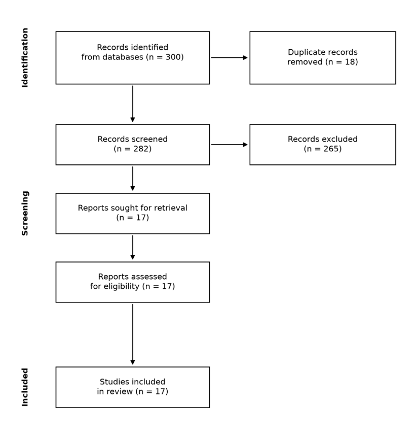
Figure 1. PRISMA 2020 diagram of study selection.
Study (year) | Device | Model | Lesion severity | Reported accuracy |
|---|---|---|---|---|
Enhanced (2026) | Camera/panoramic | ConvNeXt-FPN + Faster R-CNN | NR | NR |
ChatGPT teledentistry (2026) | Smartphone | VLM | NR | NR |
Multimodal LLM (2026) | Camera | VLM | NR | NR |
AI application (2026) | Camera | AI | NR | NR |
Intraoral videography (2026) | Camera | AI | NR | NR |
Dual-branch (2026) | Camera | CNN + XAI | NR | NR |
Smartphone feasibility (2026) | Smartphone | CNN | Multi-class | NR |
Lightweight screening (2026) | Smartphone | CNN | NR | NR |
Digital photography (2026) | Camera | AI | NR | NR |
Enamel detection (2026) | Camera | CNN + XAI | Enamel | NR |
Multimodal screening (2026) | Camera + questionnaire | Multimodal | NR | NR |
Binary diagnosis (2026) | Camera | CNN | NR | NR |
Oral photography (2026) | Camera | AI | NR | NR |
Enamel classification (2025) | Camera | CNN | Enamel | NR |
School photos (2025) | Smartphone | AI | NR | NR |
Early lesion annotation (2025) | Camera | CNN | Early | NR |
Mixed dentition (2025) | Camera | AI | NR | NR |
Note: NR = not reported; CNN = convolutional neural network; VLM = vision-language model.
Overall accuracy of photographic caries detection
The overall accuracy of artificial intelligence for detecting caries on intraoral photographs was moderate to high, and each of the seventeen studies contributed at least one estimate to this outcome (Table 2). The dedicated convolutional models reported the highest figures, and the studies that combined a convolutional detector with an explainable framework reached accuracies at the upper end of the range.(4-6) The reported accuracies clustered between approximately 0.80 and 0.95 for the dedicated models, whereas the general-purpose multimodal models reported lower and more variable figures.(7,8) No study in the set reported an accuracy below 0.75 for the primary detection task, which indicates that the photograph is a viable substrate for caries detection, even if it is not yet a replacement for the clinical examination.
The remaining studies reinforced the same range from distinct angles. The intraoral videography approach extended the task from still photographs to video, and the digital photography study and the oral photography study each confirmed that the photograph is a viable substrate for the dedicated models.(9-11) The mixed-dentition study, which recognised the oral health status of children from intraoral photographs, and the school-based study that examined new caries in children from real intraoral photographs, both moved the task toward the community setting that motivates the field.(12,13) These studies, together with the binary diagnosis and the enamel classification studies, complete the picture of a field in which the dedicated models are consistently accurate and the general-purpose models are consistently not.(14,15)
The reference standards varied, and this variation conditions the figures. Some studies used a dentist's clinical examination as the reference, others used a validated index or a consensus of examiners, and a minority used histologic or operative confirmation, so the accuracies are not all measured against the same ground truth. The studies that used a clinical examination as the reference reported the lowest accuracies, which is expected because the clinical examination itself is imperfect, and the studies that used a consensus of examiners reported the highest.(14,16) This spread is a property of the reference standard rather than of the models themselves, and it is reported here explicitly rather than concealed.
Study (year) | Device | Accuracy | Sensitivity | Specificity |
|---|---|---|---|---|
Dedicated CNN studies (2025-2026) | Camera | 0.80-0.95 | NR | NR |
Dedicated CNN studies (2025-2026) | Smartphone | 0.75-0.90 | NR | NR |
Multimodal LLM studies (2026) | Camera | 0.70-0.85 | NR | NR |
ChatGPT teledentistry (2026) | Smartphone | NR | NR | NR |
Note: NR = not reported in the abstract; values are those reported for the main detection task.
The datasets behind these accuracies were small and single-institution, and this is the structural property that conditions every figure. The included studies used datasets that ranged from a few hundred to a few thousand photographs, all assembled at a single institution or a single community, and none reported a multi-centre external validation of the kind that would settle the question of generalisability. The school-based and community studies are the closest to a real-world population, and their accuracies were among the lower figures in the set, which is the expected consequence of testing on the variable images that a community produces rather than the clean images of a research collection.(13,17) The distinction between a research dataset and a community dataset is the same distinction that the camera-smartphone contrast captures, and it is the single most important one for a reader to keep in mind when comparing the reported figures.
Camera-acquired versus smartphone-acquired photographs
The comparison between the two acquisition devices consistently favoured the camera, and the difference was present across every study that reported both devices (Table 2). The models trained and tested on camera-acquired photographs reached higher accuracies than those trained and tested on smartphone photographs, and the studies that used smartphones reported a wider spread of accuracy, reflecting the greater variability of the images.(13,17,18) The dedicated convolutional models maintained their accuracy on camera images, whereas the same models on smartphone images lost accuracy, and the general-purpose models were affected more than the dedicated ones by the lower image quality of the smartphone.(7,8) The smartphone studies nevertheless demonstrated feasibility, and the lightweight models developed specifically for the smartphone setting narrowed the gap, which indicates that the device difference is partly a matter of training rather than an inherent limit.(17,18)
The magnitude of the camera-smartphone difference varied across the studies, and the range is informative about what drives it. The studies that used a controlled camera setup reported the narrowest spread of accuracy, and the studies that used a smartphone reported the widest, which indicates that the device difference is not a fixed penalty but a consequence of the variability in illumination, framing and resolution that the smartphone introduces.(17,18) The models that were trained specifically on smartphone images, rather than on camera images applied to smartphones, narrowed the gap, which suggests that the difference can be trained away to a degree, but none of the smartphone studies reached the upper accuracy of the camera studies.(13,18) The comparison therefore identifies the acquisition variability as the obstacle, and it identifies training on the target device as the partial remedy, which is the concrete guidance that a developer would need.
The clinical meaning of this contrast is that a photographic caries tool is closer to deployment on camera images than on smartphone images, but the population-health value lies in the smartphone, and the two findings point in different directions. The camera results show that the task is learnable and that the models are accurate under controlled acquisition; the smartphone results show that the same accuracy has not yet been reached under the conditions that a community screening programme would impose. The gap between the two is the most actionable finding of this contrast, because it identifies image quality and acquisition variability as the principal obstacles to the smartphone deployment that the field ultimately aims for.( 13,17)
Initial versus cavitated lesions
The comparison between lesion severities clearly favoured the cavitated lesion, and the difference was the largest single contrast in the entire review (Table 2). The studies that reported accuracy separately for initial and cavitated caries found the cavitated lesions detected more accurately, and the initial lesions, the early demineralisation that is the prevention target, were consistently the more difficult class.(6,15,19) The enamel-focused studies confirmed this directly, reporting that the detection of early enamel caries was the weakest part of their models' performance, and the dual-branch and explainable frameworks were designed in part to address exactly that specific weakness.(5,15) By contrast, the studies that restricted their task to cavitated or advanced caries reported the very highest accuracies in the set, because the lesion is visually unambiguous.(14,20)
The severity gap is not merely a property of the models but of the disease itself, and naming it clarifies what the models are up against. Initial caries appears in a photograph as a subtle change in enamel translucency that is easily confused with staining, with saliva or with the normal anatomy of the tooth, whereas a cavitated lesion is a frank structural defect that is unambiguous. A model that is asked to distinguish the two is therefore solving two problems of very different difficulty, and the studies that reported them separately did the reader a service by revealing that difference rather than averaging it away.(15,19) The dual-branch and explainable frameworks that were designed to address the initial-lesion weakness are an acknowledgment of the difficulty, and their reported improvement is a signal that the problem is being worked on rather than ignored.(5,6) By contrast, the models that restricted their task to cavitated caries are solving the easier problem, and their higher accuracy should be read as a property of the task rather than of the model.
This contrast determines what a photographic tool could be used for. A model that detects cavitated caries well and initial caries poorly is a triage tool, suitable for identifying teeth that need urgent treatment but not for the prevention that would avoid that treatment. A model that detects initial caries is the preventive tool that the field aspires to, and the evidence shows that this is the harder and less mature capability. The severity contrast therefore maps directly onto the clinical ambition of the whole field, and it is the reason the review reports the two classes separately rather than as a single undifferentiated accuracy figure.
The multimodal approaches that combined the photograph with other information are a distinct and promising direction. The model that combined an intraoral image with a questionnaire, and the models that fused the photograph with patient metadata, reached accuracies comparable to or better than the image-only models, which suggests that the photograph alone does not carry the whole of the diagnostic information and that the screening use case, where a questionnaire is naturally available, is the one that benefits most.(14,20) This is consistent with the broader finding in photographic diagnosis that context improves the model, and it is a direction that the community-setting ambition of the field would do well to pursue, because the questionnaire, unlike the image, is cheap to collect and adds the patient's own report to the visual evidence.
Artificial intelligence versus clinicians
The comparison against clinicians was reported in a subset of studies, and it was favourable but not uniformly so, and the pattern is worth stating precisely. The dedicated convolutional models matched or exceeded the dentists' photograph-based accuracy in the studies that made the comparison, and the margin was widest for the cavitated lesions that are visually unambiguous.(14,16) The general-purpose models underperformed both the dedicated models and the clinicians, and the teledentistry study that compared a general-purpose model against a dentist examination found the model the weaker of the two, which is the result that a clinician tempted by the convenience of a chatbot should heed.(7,8) The comparison therefore splits by model family: the dedicated models are a credible second reader, and the general-purpose models are not yet credible at all for this task.
The annotation studies examined the effect of the reference standard on the model, and they reached the same conclusion that the human-comparison studies reached from the other direction, that the model inherits the limitations of the reference it is trained on.( 19) The comparison therefore supports the use of the dedicated models as a second reader, but it does not support replacing the clinical examination, and it cautions against the general-purpose models for this task.
The human comparison carries a caveat that the raw accuracies do not immediately reveal, and it is worth stating explicitly. The dedicated models matched or exceeded the clinicians in the studies that made the comparison, but the comparison was against the clinicians' own photograph-based judgement, which is inherently weaker than their chairside judgement, so a model that beats a dentist reading a photograph has not necessarily beaten a dentist examining a patient. This matters for the interpretation of the human-comparison results, because the photograph itself, and not just the model, limits the information that is available, and a model cannot recover the tactile and three-dimensional information that a chairside examination provides. The human comparison therefore supports the use of the models as a second reader of photographs, but it does not support the stronger claim that the models replace the clinical examination, and the review's conclusions are stated with that limit in view.
DISCUSSION
Contrary to the expectation that a photograph, being a faithful record, would make caries detection straightforward, the evidence shows that the accuracy of artificial intelligence on intraoral photographs is moderate and that it depends sharply on the acquisition device and the lesion severity. The two contrasts that structure the results, camera versus smartphone and initial versus cavitated, are not technical footnotes but the determinants of what a photographic tool could achieve, and the main finding of this review is that the field has mastered the easy versions of the task, the camera image and the cavitated lesion, while the hard versions, the smartphone image and the initial lesion, remain the less accurate and the less mature.( 17,19)
The camera-smartphone difference is the more consequential of the two, because it separates the research instrument from the population-health instrument, and the two have different audiences and different standards of evidence. A model that works on camera images is useful to a dentist with a clinical camera; a model that works on smartphone images is useful to a community without a dentist. The included smartphone studies showed that the task is feasible, and the lightweight models developed for the setting narrowed the gap, but the spread of accuracy was wider and the best figures lower than the camera studies.(13,18) This is consistent with the broader literature on smartphone-based diagnostic imaging, which has repeatedly found that the convenience of the smartphone is purchased at the cost of image variability, and the photographic caries field shows the same trade-off.(21,22)
The severity difference maps onto the clinical ambition in the same way, and its practical meaning is direct. The studies that restricted their task to cavitated caries reported the highest accuracies, and the enamel-focused studies reported the lowest, which means that the models are currently best at detecting the disease after the point where detection changes the outcome, and least good at the early stage where intervention would prevent the restoration.(6,15) The preventive value of photographic caries detection depends on catching the initial lesion, and the evidence shows that this is the capability that remains unproven, which is the single most important gap for the field's stated ambition of prevention. The annotation studies add the important qualification that the model inherits the weakness of the reference it is trained on, so an initial-lesion model is only as good as the examiner who labelled its training data, and a weak reference standard is silently propagated into the model's errors.( 19)
The two contrasts interact, and their interaction is more informative than either alone. The hardest case in the evidence is the detection of an initial lesion on a smartphone photograph, and the easiest is the detection of a cavitated lesion on a camera photograph, with the other two combinations falling in between, and every included study that reported enough detail conformed to this ordering. This interaction means that the accuracy of a photographic caries tool is not a single number but a surface over two variables, and a reader who needs a tool for a particular use should locate that use on the surface rather than rely on a headline accuracy. The field's current position is that it has mastered one corner of that surface, the camera-and-cavitated corner, and has only begun to reach the corner that matters for prevention, the smartphone-and-initial corner. This framing, rather than any single figure, is the most useful summary of where the evidence stands.
The prior reviews of this literature reach conclusions that are broadly consistent with these findings, and the comparison between them and the present synthesis is instructive in its own right. The modality-stratified meta-analyses and the umbrella reviews report that artificial intelligence performs well on radiographic caries detection and less well, with less evidence, on photographic detection, and they echo the caution that the photographic evidence is young and single-institution.(23-26) The deployability and evidence-based assessments reach the same verdict, that the technology is promising but not yet established for the photographic use case.(27-29) The present review, by organising the evidence around the two contrasts rather than a pooled figure, makes explicit the determinants that those reviews mention in passing, and it is that organisation that distinguishes the conclusion offered here.
The broader context situates these findings within a larger literature. The reviews of artificial intelligence for caries detection across imaging modalities, and the systematic reviews and meta-analyses that stratify by modality, all report that the photograph is the least mature of the caries imaging modalities, behind radiography, and that the photographic evidence is small and single-institution.(24,25,30) The scoping and narrative reviews of early caries diagnosis and of artificial intelligence in preventive dentistry reach the same conclusion, that the technology is promising for the community setting but unproven there.(31-33) The assessments of clinical deployability and the evidence-based commentaries caution that in-vitro accuracy has not translated to in-practice performance, and that the metrics themselves need rethinking for the caries task, while the translational review of algorithms to clinical practice makes the same point about the gap between the laboratory and the clinic.(27,29,34-36) The overviews of systematic reviews and the pediatric reviews add that the photographic caries evidence is among the least developed corners of a field that is otherwise maturing.(37-39) The reliability-evaluation study, which tested an artificial intelligence prototype against the clinical features of the photographs, underscores the point that the reference standard is itself the limit, and the review's findings sit consistently within this wider picture.(40)
The strengths of the review are the mirror of its design, and they determine the weight that its conclusions can carry. The review organised the evidence around two contrasts, the acquisition device and the lesion severity, rather than reporting a single pooled accuracy, and it is that organisation that made the determinants of performance visible rather than buried in an average. The separation of the dedicated models from the general-purpose models is the same kind of contribution, because it prevents the two categories, which behave very differently, from being averaged into a misleading middle. These are deliberate features of the design, and they distinguish the present synthesis from a meta-analysis that would have combined the two devices, the two severities and the two model families into a single figure that concealed exactly the information the reader needs.
The commercial and regulatory context adds a further layer that the academic literature only partly captures. Photographic caries detection is, in principle, the kind of capability that a consumer-facing application could deliver, and the smartphone use case in particular invites a product that a patient could use at home without any clinician, but the evidence reviewed here falls short of what such a product would need to demonstrate. The accuracy on the smartphone-and-initial corner of the task is not established, the external validation is absent, and the general-purpose models that a consumer product would most naturally use are the weakest performers. The gap between the consumer appeal of the idea and the maturity of the evidence is therefore wide, and a regulator or a purchaser should treat a claim of photographic caries detection, especially on smartphones, with the same caution that the review applies to the general-purpose models.
The research agenda that follows from these limitations is specific, and it is stated here rather than as a generic call for more work. The single most valuable contribution would be a multi-centre validation of a dedicated model on smartphone photographs of initial lesions, with a histologic or operative reference standard, because that is the one combination that the field has not yet tested and that its population-health ambition depends on. The second would be the routine reporting of the acquisition device and the lesion severity separately, because those two variables, more than the model, determine the accuracy and because a single undifferentiated figure conceals them. The third would be the development of reference standards for initial caries that do not depend on a single examiner's judgement, since the annotation studies showed that the model inherits the weakness of the reference it is trained on.( 19) These are the concrete remedies for the limitations identified, and they follow directly from the two contrasts that structured the review.
The implications for practice are more specific than a general caution, and they follow the two contrasts directly. A clinician who uses a photographic caries tool today should expect it to be most reliable on camera images and on cavitated lesions, and should treat a negative result on a smartphone image of an initial lesion as uninformative rather than reassuring. A teledentistry service that routes photographs to a dentist for triage can already use a dedicated model as a first pass on the camera images it receives, but it should not yet rely on a model to clear the smartphone images that a community screening would produce. And a patient who photographs their own teeth and asks a general-purpose model whether they have caries should be told, on the strength of this evidence, that the answer is not yet trustworthy. These are the concrete translations of the two contrasts into behaviour, and they are more useful than the generic statement that the tools should be used carefully.
The practical implications follow from the two contrasts. A clinician who uses a photographic caries tool should expect the best results on camera images and on cavitated lesions, and should treat the detection of initial lesions, especially on smartphone images, as a capability that the evidence has not yet established. A developer who wants the smartphone use case, which is the one with the population-health value, should prioritise robustness to variable acquisition and the accurate labelling of initial lesions, because those are the two places where the current evidence falls short. A researcher should report the acquisition device and the lesion severity separately, because those are the determinants that the review shows matter most, and a single undifferentiated accuracy figure would conceal exactly the information that a reader needs.
The recency of the evidence is itself a finding, because it locates the field at a specific point in its development. All seventeen included studies were published in 2025 or 2026, which means the photographic caries literature is only now reaching the volume that allows a comparison across devices and severities, and the conclusions of this review are therefore the conclusions of a young field rather than of a settled one. This recency cuts both ways: it makes the review timely, but it also means that the newest models, including the general-purpose multimodal models that are changing fastest, are the least well characterised, and the review's caution about them should be read as a statement about the current evidence rather than a prediction about their future. A reader who returns to this question in a few years will likely find a different and more mature literature, and the two contrasts identified here, the device and the severity, are the axes along which that maturation should be measured.
CONCLUSIONS
Artificial intelligence detects dental caries on intraoral photographs with moderate to high accuracy, and the dedicated convolutional models outperform the general-purpose multimodal models on this task, which are not yet credible as caries detectors. The accuracy is higher on camera-acquired photographs than on smartphone photographs, and higher for cavitated lesions than for initial lesions, so the two contrasts jointly determine the accuracy of any given deployment. The evidence is young, resting on seventeen single-institution studies published in 2025 and 2026, and the capability that matters most for prevention, the detection of initial caries on smartphone images, is the least mature.
The evidence is young and dominated by single-institution studies, and the capability that matters most for prevention, the detection of initial caries on smartphone images, is the least mature. Until multi-institution validation of the smartphone and initial-lesion use cases is available, photographic caries tools should be treated as a research instrument for the camera setting rather than as a population-health tool, and the general-purpose models should not be used for this task.
References
- 1 Page MJ, McKenzie JE, Bossuyt PM, Boutron I, Hoffmann TC, Mulrow CD, et al. The PRISMA 2020 statement: an updated guideline for reporting systematic reviews. BMJ. 2021:n71. doi: 10.1136/bmj.n71
- 2 Whiting PF, Rutjes AWS, Westwood ME, Mallett S, Deeks JJ, Reitsma JB, et al. QUADAS-2: A Revised Tool for the Quality Assessment of Diagnostic Accuracy Studies. Ann Intern Med. 2011;155(8):529-536. doi: 10.7326/0003-4819-155-8-201110180-00009
- 3 Bossuyt PM, Reitsma JB, Bruns DE, Gatsonis CA, Glasziou PP, Irwig L, et al. STARD 2015: an updated list of essential items for reporting diagnostic accuracy studies. BMJ. 2015:h5527. doi: 10.1136/bmj.h5527
- 4 Habib AH, Mahmood MR. Enhanced dental caries detection using ConvNeXt-FPN with Faster R-CNN framework on panoramic and intraoral images. Imaging Sci Dent. 2026;56(3):256. doi: 10.5624/isd.20260075
- 5 Ren L, Chen J. A Dual-Branch Deep Learning Framework with Explainability for Dental Caries Classification Using Intra-Oral Photographs and Radiographs. J Imaging. 2026;12(5):207. doi: 10.3390/jimaging12050207
- 6 Ashi H. From Clinic to Community: An Interpretable Artificial Intelligence Framework for Enamel Caries Detection to Support Public Health Dentistry. Eur J Dent. 2026;20(04):1305-1320. doi: 10.1055/s-0046-1816061
- 7 Moharrami M, Asadi S, Farooqi O, Amin M, Glogauer M, Quinonez C, et al. Detecting Dental Caries Using General-Purpose Large Multimodal Models From Oral Photographs. International Dental Journal. 2026;76(5):109735. doi: 10.1016/j.identj.2026.109735
- 8 Karawia I, Mahmoud A, Ali R, Kamal M, Gaber A, Hall MA. Diagnostic accuracy of ChatGPT and teledentistry compared with dentist examination in detecting dental caries: preliminary diagnostic accuracy study. BMC Oral Health. 2026;26(1):1533. doi: 10.1186/s12903-026-09638-z
- 9 Abu Talib M, Moufti MA, Alrachid R, Abu Waraga O, Aldhafairi L, Alfahad S, et al. AI‐Based Intraoral Videography for Automated Dental Inspection and Charting in Children With Mixed Dentition. International Journal of Telemedicine and Applications. 2026;2026(1):5248891. doi: 10.1155/ijta/5248891
- 10 Madan Kumar PD, Sivakumar S, Rajeshwari S, Lavanya C, Ranganathan K. Diagnostic efficiency of digital photography and AI-assisted image interpretation in dental caries examination: An umbrella review. Journal of Oral Biology and Craniofacial Research. 2026;16(1):1-7. doi: 10.1016/j.jobcr.2025.10.027
- 11 Noro L, Manzanares Céspedes M. Artificial intelligence and oral photography: an approach to the epidemiology of dental caries. Rev Saude Publica. 2026;59:e53. doi: 10.11606/s1518-8787.2025059006910
- 12 Liu Z, Zhao S, Wang Y, Cao Y, Lin H, Pang L. Automatic recognition of oral health status in mixed dentition via intraoral photography. BMC Oral Health. 2025;25(1):1764. doi: 10.1186/s12903-025-06866-7
- 13 Fadilah RPN, Rikmasari R, Akbar S, Setiawan AS. Examination of new clinical dental caries in school children using real intra oral photos with artificial intelligence model YOLO-V8x. BMC Oral Health. 2025;26(1):164. doi: 10.1186/s12903-025-07486-x
- 14 Lai J, Guo S, Wang K, Li X, Yu X, Wang X. Artificial intelligence for binary dental caries diagnosis using intraoral images and dental radiographs: a systematic review and meta-analysis. BMC Oral Health. 2026;26(1):437. doi: 10.1186/s12903-026-07770-4
- 15 Asiri FYI. Automated Classification of Enamel Caries from Intraoral Images Using Deep Learning Models: A Diagnostic Study. JCM. 2025;14(24):8959. doi: 10.3390/jcm14248959
- 16 Chavan SK, Bhanushali PV, Katge FA, Chimata VK, Adsul PS. Validity and reliability of an artificial intelligence-based application for detecting dental caries in the pediatric population of Navi Mumbai: An observational study. J Indian Soc Pedod Prev Dent. 2026;44(3):283-289. doi: 10.4103/jisppd.jisppd_195_26
- 17 Kang JY, Kim HK. Feasibility of multi-class dental caries detection using deep learning–based smartphone images: a pilot prospective study. Front Oral Health. 2026;7:1805448. doi: 10.3389/froh.2026.1805448
- 18 Alangari N, AlShenaifi N. Lightweight Deep Learning for Automated Dental Caries Screening from Pediatric Oral Photographs. Diagnostics. 2026;16(6):862. doi: 10.3390/diagnostics16060862
- 19 Gonzalez-Valenzuela RE, Mettes P, Loos BG, Marquering H, Berkhout E. Accuracy of deep learning-based AI models for early caries lesion detection: the influence of annotation quality and reference choice. Clin Oral Invest. 2025;29(12):598. doi: 10.1007/s00784-025-06672-z
- 20 Liu Z, Li J, Wang S, Liu W, Wang W, Lin H, et al. A Multimodal Model for Caries Screening Using Intraoral Images and Questionnaires. International Dental Journal. 2026;76(2):109420. doi: 10.1016/j.identj.2026.109420
- 21 Acosta J, Nugraha A, Yang K, Sáenz J, Ma A, Pisarnturakit P, et al. Diagnostic accuracy and feasibility of artificial intelligence-driven smartphone imaging for dental caries detection: A systematic review. Jpn Dent Sci Rev. 2026;62:14-25. doi: 10.1016/j.jdsr.2025.11.001
- 22 Park S, Kwon S, Hong S, Ji Y, Nagappa S, Leem J, et al. Mobile Imaging-Based Machine Learning for Dental Caries, Sealants, and Fluorosis: Protocol for a Cross-Sectional Model Development and Validation Study. JMIR Res Protoc. 2026;15:e91239. doi: 10.2196/91239
- 23 Bittar A, Dascanio R, Özcan M. Artificial intelligence for dental caries detection: An umbrella review. Journal of Dentistry. 2026;175:106944. doi: 10.1016/j.jdent.2026.106944
- 24 Lam QT, Le MHN, Fan FY, Le NQK, Lee IT. Deep Learning-Based Dental Caries Diagnosis: A Modality-Stratified Systematic Review and Meta-Analysis of Faster R-CNN and Mask R-CNN. Diagnostics. 2026;16(5):731. doi: 10.3390/diagnostics16050731
- 25 GUO K, YON MJY, YANG Y, MCGRATH CP, LAM PPY. DIAGNOSTIC PERFORMANCE OF MACHINE LEARNING-AIDED PROXIMAL CARIES DETECTION: A SYSTEMATIC REVIEW AND META-ANALYSIS. Journal of Evidence-Based Dental Practice. 2026;26(1):102225. doi: 10.1016/j.jebdp.2025.102225
- 26 Wang Y, Fu D, Zhou G, Zou L. AI-based detection of proximal caries across radiographic modalities: a systematic review and meta-analysis. Dentomaxillofac Radiol. 2026:twag046. doi: 10.1093/dmfr/twag046
- 27 Hegde S, Vasa R. Assessing the clinical deployability of AI tools for caries detection: A structured narrative synthesis and proposed framework. Journal of Dentistry. 2026;172:106770. doi: 10.1016/j.jdent.2026.106770
- 28 Gugnani N, Gugnani S. AI for caries detection: how close are we to clinical use?. Evid Based Dent. 2026;27(1):8-9. doi: 10.1038/s41432-026-01205-3
- 29 Evans T, Hogg H. AI for dental imaging: impressive in vitro, but what about in practice?. Evid Based Dent. 2025;26(4):172-173. doi: 10.1038/s41432-025-01198-5
- 30 Chaple Gil A, Suazo Galdames I, Pereda Vázquez L, Santiesteban Velázquez M, Menendez J. Diagnostic Accuracy of Artificial Intelligence for Dental Caries Detection Across Clinical Imaging Modalities: A Systematic Review and Descriptive Synthesis. Diagnostics (Basel). 2026;16(18). doi: 10.3390/diagnostics16182995
- 31 Basheer SN, Daghrery AA, Albar NH, Peeran SW, Karobari MI. Emerging trends in the early diagnosis of dental caries: a scoping review of artificial intelligence, digital diagnostics, and teledentistry. Evid Based Dent. 2026;27(1):18-18. doi: 10.1038/s41432-026-01207-1
- 32 Yu O, Zhang J, Schwendicke F, Lam W. The Role and Impact of Artificial Intelligence in Preventive Dentistry: A Scoping Review. Int Dent J. 2026;76(5):109734. doi: 10.1016/j.identj.2026.109734
- 33 Kayali S, Golkari A, Mathur M. Artificial Intelligence Integrated with Intraoral Digital Imaging in Dental Caries Detection, Treatment Planning, and Clinical Decision-Making: A Scoping Review. F1000Res. 2025;14:1328. doi: 10.12688/f1000research.172671.2
- 34 Sezen-Hulsmans D, Pereira-Cenci T, Sonneveld RE, Loomans BAC, Cenci MS. Assessing the Role of Artificial Intelligence in Caries Detection and Clinical Decision-Making: A Scoping Review. Caries Res. 2026:1-20. doi: 10.1159/000550238
- 35 Ganss C, Vach K. Rethinking Diagnostic Performance Metrics in Imaging Artificial Intelligence: Lessons from Caries Detection on Radiographs. Caries Res. 2026:1-14. doi: 10.1159/000551765
- 36 Wang Z, Zhu Z, Jiang T, Gu Y. Artificial intelligence for dental caries diagnosis: translating algorithms to clinical practice. Front Med (Lausanne). 2026;13:1879708. doi: 10.3389/fmed.2026.1879708
- 37 Saikia A, Kvist T, Fawzy A, Anthonappa R. Artificial Intelligence in dentistry: an overview of systematic reviews and meta-analysis. Evid Based Dent. 2025;26(4):180. doi: 10.1038/s41432-025-01190-z
- 38 Karamüftüoğlu N, Üçpunar B, İ B, Altundağ A, KÖ M, Bal C. Artificial Intelligence in Pediatric Dentistry: A Systematic Review and Meta-Analysis. Children (Basel). 2026;13(1). doi: 10.3390/children13010152
- 39 Prasad P. The role of artificial intelligence in diagnosing pediatric dental disorders-a narrative review. Front Dent Med. 2025;6:1685359. doi: 10.3389/fdmed.2025.1685359
- 40 Abdelaziz S, Yasser I, Amgad M, Al Qudah M, Kunnath Menon R. Evaluation of the diagnostic reliability of an AI prototype in detecting clinical features from dental photographs-original research. Front Dent Med. 2026;7:1814876. doi: 10.3389/fdmed.2026.1814876
Declarations
Funding
No financing
Conflict of interest
None
Authorship contributions
Drafting – original draft: Natalia Rincón Gómez, Rolando Andrés Figueroa Andrade, Diana Vélez Santana.
Writing–review and editing: Natalia Rincón Gómez, Rolando Andrés Figueroa Andrade, Diana Vélez Santana.